As a veterinarian specializing in sheep reproduction, I know that understanding each ewe’s ovarian cycles is vital for flock productivity. Portable ultrasound imaging has become an essential tool on the farm. By scanning ewes’ ovaries, I can non-invasively see follicle development and corpora lutea (CL) – signs of a ewe’s current reproductive status. Modern portable ultrasound scanners are lightweight and even smartphone-compatible, allowing me to monitor reproductive organs in real time. This helps me decide when to breed, synchronize estrus, or cull non-productive animals with confidence. In the sections below, I’ll explain how we use portable ultrasound to evaluate ovarian activity in sheep and why this approach is transforming sheep breeding.

Understanding Sheep Ovarian Cycles
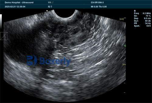
Sheep are seasonal breeders. In the breeding season, most ewes cycle roughly every 17 Jours, going through follicular growth and ovulation. During each cycle, one or more follicles grow on the ovary and, at ovulation, un corpus luteum forms on each ovary. High-resolution ultrasound can visualize this process: active follicles appear as dark (fluid-filled) circles on the scan, while a corpus luteum shows as a denser structure with a thick wall. Importantly, portable ultrasound lets us confirm puberty and cyclicity in young ewe lambs by detecting follicles ≥3 mm and identifying CLs. Par exemple, spotting a newly formed CL on ultrasound is “definitive proof of ovulation and cyclicity”. By scanning periodically, we can even track the normal wave-like pattern of follicle growth and regression in each cycle. Pratiquement, a ewe typically has several follicular waves per cycle, and portable ultrasound makes it possible to watch these changes over time without any discomfort to the animal.
Using Ultrasound for Ovarian Monitoring
In my practice, we usually perform transrectal ultrasound exams on ewes during handling or synchronization protocols. A high-frequency linear or convex probe (around 5–7.5 MHz) provides the best resolution for the small sheep ovary. Because the probe is inside the rectum, it’s very close to the ovary, yielding clear images even of tiny follicles. Transrectal scanning is completely non-invasive and quick. With training, an operator can scan an entire flock very efficiently. In fact, experienced technicians can examine up to 100–150 ewes per hour at high accuracy. We apply ultrasound gel to ensure good contact and gently move the probe until we locate each ovary. On the screen, the ovary looks like a small almond-shaped organ; we measure any visible follicles or CL. The real-time images tell us immediately whether a ewe has dominant follicles, a recent ovulation (CL), or possibly inactive ovaries.
Ultrasonic Indicators of Ovarian Activity
Portable ultrasound provides several key indicators of reproductive status:
-
Follicle detection and size. Growing follicles appear as anechoic (dark) circles. We count them and measure their diameter. A large follicle (for example > 3 mm in a ewe lamb) indicates the ewe is in the follicular phase. Tracking the largest follicle’s growth over days can predict imminent ovulation.
-
Corpus luteum (CL) detection. A CL appears as a small, bright-walled structure. Detecting one means that ovulation has occurred in the previous cycle. The CL’s size and shape can be recorded; if it is smaller or absent, the ewe may not have ovulated. Identifying CLs confirms the ewe has entered the luteal phase.
-
Doppler blood flow. Many portable scanners include Doppler mode. By adding Doppler to B-mode imaging, we can see blood flow in the ovary or CL. A functioning CL will show characteristic blood flow (sometimes called the “pavement” or “irrigation” of the CL), confirming its viability. This is useful because it links the ultrasound image to hormonal activity (a well-vascularized CL is producing progesterone). Advances in ultrasound even allow detecting subtle vascular changes around follicles and CLs.
-
Uterine checks. Although our focus is ovaries, we also glance at the uterus during scanning. Uterine health (thickness of the endometrium, presence of fluid, etc.) can also be evaluated, helping diagnose conditions that might affect fertility.
Using these observations, we can assess where each ewe is in her cycle. Par exemple, on day 0 (day of ovulation) the ovary may have a large (4–6 mm) dominant follicle. Two to three days later, a corpus luteum forms, which we spot by ultrasound. If we scan again on day 7–9, the dominant follicle will have regressed if the ewe is not pregnant, while the CL will persist (unless she was exposed to progesterone treatments). By watching these patterns, we know if and when ovulation happened.
Key Benefits of Portable Ultrasound
Portable ultrasound has several practical advantages for sheep reproduction:
-
Non-invasive and safe. It uses only harmless sound waves. No sedation or radiation is needed. This means the exam does not stress the ewe and can be repeated many times if needed.
-
Real-time quantitative data. We can measure follicle diameters, CL size, and count multiple structures right on the spot. This objective data guides precise decisions (e.g. “this ewe has a 5 mm follicle and a small CL – she’s likely just finished estrus”).
-
Rapid and efficient. A skilled operator can scan dozens of ewes in an hour, much faster than older methods. With good animal handling, we routinely screen entire groups. For pregnancy checks, some farmers report examining up to 100 ewes/hour with ~98% accuracy.
-
Immediate feedback. Unlike blood tests or waiting for behavioral signs, ultrasound shows results instantly. Par exemple, we know within seconds if a CL is present or if follicles are developing. This lets us adjust nutrition or breeding plans without delay.
-
Improved reproductive management. Having ultrasound data helps us breed smarter. We avoid breeding ewes that aren’t cycling or have abnormal ovaries, and we can time insemination or ram placement when ovulation is imminent. Over time, this data-driven approach increases conception rates and saves on feed by not overfeeding non-pregnant ewes.
-
Non-repetitive cost. Once purchased, the ultrasound is reusable every season. It replaces less reliable methods (like progesterone assays with false positives or risky palpation). In my experience, it pays for itself by preventing costly mistakes.
Together, these benefits make portable ultrasound an indispensable tool on a modern sheep farm.

Ultrasound-Guided Breeding Decisions
Pratiquement, I use ultrasound findings to make specific management decisions:
-
Ewe lamb puberty and breeding. Before first breeding, we scan ewe lambs (~7–10 months old) to confirm puberty. Ewe lambs with active follicles and/or a CL are ready for breeding, while those with inactive ovaries can be given more time to mature or culled as replacement stock. This targeted approach “maximizes first-service conception rates and reduces dystocia”. Par exemple, in New Zealand, farmers scan ewe lambs before autumn mating to ensure only the most reproductively advanced lambs are bred. This aligns lambing with peak spring grass and improves lamb survival.
-
Estrus synchronization. When using hormonal protocols (CIDR, PMSG, etc.), ultrasound helps us apply treatments effectively. We only synchronize ewes that are truly cycling; those without follicles or CLs are excluded or managed differently. By confirming ovarian status beforehand, synchronization protocols become more effective and cost-efficient. On large farms (e.g. in Australia), portable ultrasound allows quickly sorting thousands of ewes into cycling vs. non-cycling groups for synchronized breeding.
-
Nutritional flushing. In some flocks, we identify ewes with poor ovarian activity before mating. If a ewe is well-fed but shows no sign of cycle, we may subject her to a flushing diet or additional supplements. For instance, one report noted that ultrasound picked up ovarian inactivity in nutritionally stressed ewes, prompting targeted supplementation that improved their ovulation rates.
-
Pregnancy planning. While this article focuses on ovaries, it’s worth noting that ultrasound can also confirm early pregnancy. Knowing which ewes conceived by scanning 4–6 weeks after breeding (or seeing an empty uterine horn) lets us manage feed and cull non-pregnant ewes promptly.
By integrating ultrasound findings, we make smarter decisions: which ewes to breed, which need treatment, and when to expect lambing. This precision leads to better lambing rates and more uniform flocks.
Breed, Season and Nutrition Influences
Ultrasound can also reveal how genetics, season, and feeding affect reproduction:
-
Breed differences. Some breeds reach puberty earlier or have higher ovulation rates than others. Par exemple, Merino ewe lambs may show smaller follicles at puberty compared to a more prolific breed. Regular scanning helps compare breeds under the same conditions. In pedigree programs (like in the UK), breeders use ultrasound to identify genetically superior lambs with strong ovarian activity early.
-
Seasonal effects. As day length shortens (autumn), most breeds enter estrus. In winter or spring, ewes may enter anestrus. By scanning ewes outside the main season, we can document when each flock resumes cycling. For instance, scanning in spring may reveal lingering inactive ovaries despite good body condition, signaling the need for hormonal induction or targeted feeding to boost fertility.
-
Nutritional status. A well-fed ewe should have active follicles; an underfed one may show delayed or small follicles on ultrasound. We often use scanning to verify the effect of feeding programs. If ultrasound shows poor ovarian development even after improving diet, we might adjust minerals or energy content.
In all these cases, portable ultrasound gives us concrete evidence of ovarian function under real farm conditions, guiding breeding strategies tailored to each flock’s situation.
Conclusion
Today’s sheep producers can no longer rely on guesswork for reproduction. Portable ultrasound puts clear reproductive information in our hands. In my flock, scanning has become routine: we non-invasively image each ewe’s ovaries to determine whether she is cycling, ready to breed, or in need of intervention. This technology delivers real-time, precise data (confirming ovulations, counting follicles, or detecting silent heat) that has greatly improved our conception rates and reduced wasted feed on non-pregnant animals. As one expert put it, ultrasound evaluation of ovarian activity in replacement ewes “is no longer a luxury; it’s a strategic investment in flock longevity and profitability”. Indeed, around the world—from sheep farms in New Zealand to ranches in Australia—ultrasound is helping farmers align breeding with optimal conditions.
Looking ahead, portable scanners continue to evolve (new wireless probes, AI image analysis, smartphone apps). These innovations promise even more efficient scanning on the farm. For now, having this “window” into the ovary means I can make data-driven decisions every breeding season. By combining veterinary science with practical management, portable ultrasound ensures that ewes breed at the right time, improving welfare and profitability across the flock.
References: Beef and sheep reproductive experts and producers widely document the value of ultrasound. Par exemple, Merck’s Veterinary Manual (2024) emphasizes ultrasound as a “rapid, highly sensitive and very specific” test for reproductive evaluation. Extension resources from Michigan State University describe modern portable scanners that stream images to phones and goggles for easy use. Veterinary articles note that identifying follicles and CLs via ultrasound definitively confirms ovulation. Pratiquement, industry guides (e.g. by Dawei Vet and CHISON) highlight ultrasound’s non-invasive safety and its role in precision breeding. All these sources attest that portable ultrasound is now indispensable for managing sheep reproduction in a data-driven way.