Dans la gestion moderne des petits ruminants, échographie en temps réel (RTU) technology has become a critical non-invasive method for evaluating carcass composition, particularly subcutaneous fat and muscle development. Veterinary ultrasound is widely applied to measure backfat and eye muscle in sheep and goats, aiding in selection, growth monitoring, and meat quality control. This article outlines the typical ultrasound procedures, anatomical landmarks, measurement accuracy, and the importance of high-resolution probes for reliable evaluation.
Ultrasound Scanning Region and Target Muscles
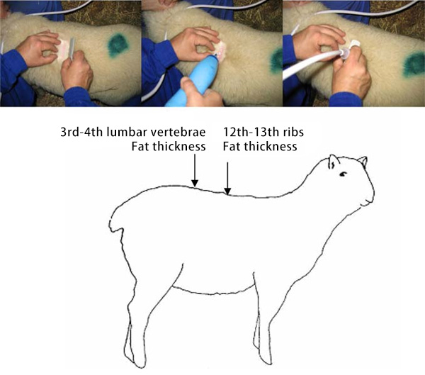
The veterinary ultrasound examination for backfat and eye muscle in sheep is typically conducted along the midline of the thoracic and lumbar regions, usually between the 12th thoracic vertebra (T12) and the 4th lumbar vertebra (L4). Anatomical orientation relies on the longissimus thoracis et lumborum (LTL) muscle and associated vertebrae.
Key parameters measured during RTU include:
-
Subcutaneous fat depth (SFD) above the LTL muscle
-
Muscle depth
Studies have shown a strong correlation (r > 0.6; P < 0.01) between these ultrasound values and corresponding carcass traits in these regions, validating ultrasound as a reliable prediction tool for meat yield and quality.
Importance of Accurate Probe Placement
Accurate probe placement is essential due to the significant variability in SFD and LTL muscle shape and thickness over short anatomical distances—both in cranio-caudal and medio-lateral directions. Inconsistent probe placement can drastically affect measurement results. Furthermore, anatomical distortions caused by animal posture and skin flexibility can also introduce discrepancies between RTU data and actual carcass measurements.
This issue is particularly pronounced in young animals, where softer skin and thinner tissue layers make it more difficult to obtain precise and repeatable readings. Donc, precision in positioning and consistent technique are vital for meaningful comparisons and longitudinal monitoring.
Ultrasound Methods for Backfat Thickness Measurement in Sheep
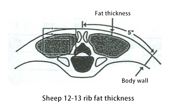
Besides the standard thoracolumbar region, veterinary ultrasound is also used to evaluate subcutaneous fat thickness in other locations, comme:
-
The sternum
-
GR site (Grade Rule measurement): Located laterally 11 cm from the spine between the 11th and 12th ribs.
The sternum region is especially suitable for assessing fat depth in goats, where subcutaneous fat tends to be thinner and more challenging to evaluate at the thoracic and lumbar sites. This difference highlights the necessity of species-specific scanning protocols.
Optimal Scanning in High-Definition Sheep Backfat Evaluation
Within the same species and between different species, fatter animals show more consistent and accurate subcutaneous fat depth measurements. Conversely, in lambs, ultrasonographic evaluation is more challenging due to the extremely thin fat layers, which limits SFD assessment.
A practical solution for this issue is the use of Sondes haute fréquence, comme 7.5 MHz transducers, which offer:
-
Higher image resolution
-
Lower penetration depth, ideal for surface-level tissues
The first 6 mm from the skin surface, which includes both the skin and subcutaneous fat, is of greatest interest. Ainsi, precise identification of the skin-fat interface is essential, given that fat thickness often measures just a few millimeters.
Resolution and Measurement Accuracy
Accurate depth measurements depend on clear visualization of tissue boundaries and interfaces. Most Appareils à ultrasons vétérinaires feature internal measurement systems with a resolution of 1 mm. Toutefois, this resolution may be insufficient when examining lean animals or when tracking small changes in tissue development over time.
To overcome this limitation, researchers have used video capture and image analysis software in conjunction with ultrasound devices to evaluate fat and muscle thickness in sheep. Compared to direct on-screen measurement, their approach offers superior repeatability and precision.
The key differences lie in resolution:
-
1 mm resolution on ultrasound monitors
-
0.1 mm resolution with image analysis software
-
0.2 mm resolution avec 7.5 MHz high-frequency probes
Such enhancements allow for the detection of subtle differences in SFD between animals, reinforcing the value of recording ultrasound images for later analysis.
Advantages of Image Recording and Post-Processing
Recording ultrasound images and performing subsequent analysis offers several significant benefits:
-
Shorter examination time per animal – beneficial in large flocks
-
Multiple measurements from the same image, including irregular zones
-
Enhanced repeatability, as interpretation of recorded images is often more accurate than real-time acquisition
These factors provide a compelling argument for integrating video documentation and software-assisted image analysis into ultrasound-based meat quality assessment protocols in sheep and goat farming.
Conclusion
Veterinary ultrasound offers a highly effective, non-invasive method for evaluating backfat and muscle quality in small ruminants. When used with high-resolution probes and supported by advanced image analysis tools, it provides reliable data that closely correlates with carcass measurements. Accurate probe placement, species-specific protocols, and post-processing analysis are essential for maximizing the accuracy and utility of ultrasound data. As demand grows for precision livestock farming, veterinary ultrasound continues to play a critical role in improving genetic selection, feeding strategies, and meat quality in sheep and goat production systems.