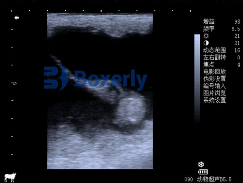
As a professional in veterinary medicine or canine reproduction, understanding the intricate changes that occur during a dog’s pregnancy is crucial for ensuring the health of both the mother and her developing puppies. Among the many diagnostic tools available, échographie vétérinaire (B-mode ultrasound) has revolutionized how we monitor canine gestation, especially between 30 et 50 days of pregnancy—a critical window marking major embryonic and fetal development milestones.

This article will provide an in-depth overview of how veterinary ultrasound monitors embryo changes during this gestational period, highlighting key morphological transformations, physiological markers such as fetal heart rate and movement, and placental development. The discussion incorporates both traditional knowledge from Chinese veterinary practice and the latest international research and clinical applications, making this resource valuable for veterinarians, breeders, and canine enthusiasts worldwide.
The Importance of Veterinary Ultrasound in Canine Pregnancy Monitoring
Pregnancy diagnosis and monitoring in dogs can be challenging due to their relatively long gestation period (environ 63 Jours) and the subtle external signs of fetal development. While palpation and hormonal assays offer some information, ultrasound provides a non-invasive, real-time imaging method that enables direct visualization of the embryos and their physiological status.
Internationally, veterinary ultrasound is considered the gold standard for confirming pregnancy after about 20-25 days post-breeding and monitoring fetal health throughout gestation. From approximately 30 days onwards, ultrasound images become increasingly detailed and informative, allowing clinicians to:
-
Identify embryo viability via heartbeats and movement
-
Observe morphological development stages
-
Detect placental growth and health
-
Predict litter size and detect fetal abnormalities early
By focusing on the 30 À 50 days pregnancy window, this article highlights a stage when the embryo transforms from a relatively simple structure into a rapidly developing fetus with recognizable anatomy, making ultrasound findings crucial for clinical decisions and reproductive management.

Embryonic Changes on Veterinary Ultrasound Around 30 Days Pregnancy
At roughly 30 days of gestation, the canine embryo undergoes significant structural development visible on ultrasound. According to research and clinical observations, including those from Chinese veterinary ultrasound monitoring, the following changes are noted:
-
From Light Points to Dense Fetal Mass: Initially, embryos appear as scattered bright points or linear echoes on the ultrasound screen. By day 30, these points consolidate into unevenly dense fetal masses or clumps, indicating tissue differentiation.
-
Distinct Head and Limb Outlines: Although still faint, the embryo’s head appears relatively large and round, while the limbs manifest as thin lines or shadows. Overall, the fetal shape on ultrasound resembles an “L” configuration or grouped clumps.
-
Umbilical Cord Visualization: A distinct umbilical cord can be seen connecting the fetus to the placenta, with its diameter increasing as the fetus grows, indicating active nutrient and oxygen exchange.
-
Heartbeat Detection: The fetal heart begins to beat visibly on ultrasound as intermittent flashes or flickers inside the thoracic cavity. The heart rate can be measured, providing important information about fetal viability.
Western veterinary literature echoes these observations, emphasizing that fetal heart rate monitoring at this stage serves as a vital sign of embryo health. According to “Canine Reproduction and Neonatology” (Johnston et al., 2019), the fetal heartbeat at 30 days ranges between 120-180 beats per minute, detectable via ultrasound Doppler or M-mode imaging.
Embryonic and Fetal Development from 31 À 35 Days
Between 31 et 35 Jours, the embryo continues morphing rapidly, as ultrasound images document:
-
Body Thickening and Morphological Change: The fetus’ body thickens progressively. The head-to-body ratio decreases as the head bends towards the body. The embryo takes on a characteristic “C” shape, with visible neck and back curvature.
-
Clear Cranial Structure: The skull bones start producing strong echoes, appearing as bright linear shadows on ultrasound. Inside the head, darker low-echo areas denote brain cavities forming.
-
Fetal Movement Begins: Sporadic fetal movements emerge and can be observed as subtle shifts or twitches on the ultrasound screen. Movement frequency generally slows down as pregnancy progresses but is crucial for assessing fetal wellbeing.
-
Placental Thickening: The placental layer shows noticeable thickening and growth, especially on the uterine wall side. This increase facilitates better fetal nourishment.
These ultrasound findings align with the clinical practices in Western veterinary obstetrics. Dr. Lisa M. Freeman, a veterinary nutritionist and researcher, emphasizes that fetal movement at mid-gestation is a reliable sign of viability, and reduction or absence of movement can indicate distress or fetal death.
Embryo and Fetal Changes Between 40 À 50 Days
From 40 À 50 days of pregnancy, the fetus enters a phase of rapid growth and organogenesis that can be visualized vividly on ultrasound:
-
Limb Bud Formation: Around 40 Jours, limb buds become apparent, and the earlier “C” shape disappears as the fetus stretches out and adopts a more natural posture resembling a newborn puppy.
-
Skeletal Structures Development: By day 43, the vertebral column and ribs become distinctly visible as strong echogenic lines. The spinal segments and their articulation points can be identified, reflecting vertebral development.
-
Internal Organs Development: Multiple low-echo dark areas in the fetal body expand quickly, representing internal organs like the liver, Reins, and intestines undergoing maturation.
-
Spinal Cord and Vertebrae Visualization: At 45 Jours, the vertebrae reflect ultrasound waves strongly, allowing clear segmentation to be seen, along with a central sagittal canal corresponding to the spinal cord.
-
Bone Formation Completion: By about day 50, the fetal rib cage is sharply defined, indicating the bone matrix formation is nearly complete.
These ultrasound features match with global veterinary findings. Par exemple, studies published in the Journal of Small Animal Practice highlight that advanced fetal skeletal imaging from 40 days onward helps predict puppy viability and timing of delivery.
Placental Development and Ultrasonographic Characteristics
The placenta plays a vital role in supporting the growing fetus, and its monitoring via ultrasound is essential. In breeds like the Golden Retriever, around day 31, a flattened, oval-shaped placental layer appears on one side of the uterine wall. This region shows homogenous weak echo signals corresponding to the placental band, which thickens progressively until about day 45. Beyond this point, placental size stabilizes.
The yolk sac—visible between days 25 and 28—adheres closely to the embryo and placenta. Ultrasound images display it as a U-shaped linear structure that transitions into a tubular shape as gestation advances. Around 27-31 Jours, the allantoic membrane appears as a weak echo boundary enveloping the embryo and yolk sac.
Western researchers concur that careful placental ultrasonography allows early detection of placental insufficiency, a major cause of fetal loss. According to “Veterinary Obstetrics and Reproduction” by Noakes et al., placental thickness and blood flow studies using Doppler ultrasound are standard practice in canine pregnancy management globally.
Practical Implications of Ultrasound Monitoring in Canine Pregnancy
The detailed ultrasound observations from 30 À 50 days pregnancy are not merely academic but have direct applications in veterinary clinical practice and dog breeding management worldwide:
-
Pregnancy Confirmation and Dating: Confirming pregnancy status and accurately dating gestation are vital for scheduling prenatal care and planning for whelping.
-
Assessment of Fetal Viability: Heartbeat monitoring and fetal movement detection allow early intervention if fetal demise is suspected.
-
Predicting Litter Size: Ultrasound can count embryos or fetuses, helping breeders prepare adequately for delivery and neonatal care.
-
Detecting Abnormalities: Early detection of fetal malformations or placental issues enables timely medical or surgical decisions.
-
Optimizing Maternal Health: Monitoring placental development guides nutritional and health management for the pregnant bitch.
These uses are embraced globally. Veterinary reproductive specialists in Europe and North America routinely employ serial ultrasounds at this gestational stage to reduce pregnancy complications and improve neonatal survival rates.
Conclusion
Veterinary ultrasound monitoring between 30 et 50 days of pregnancy provides an invaluable window into the embryonic and fetal development of the dog. From initial detection of fetal heartbeat and umbilical cord visualization to advanced skeletal and organ imaging, this technology informs clinical decision-making and enhances reproductive success.
Integrating traditional ultrasound findings with international veterinary research and standards enriches our understanding and application of this vital tool. As ultrasound technology continues advancing, including 3D and Doppler modalities, our ability to ensure healthy canine pregnancies will only improve.
For veterinarians, breeders, and canine lovers worldwide, embracing ultrasound monitoring during this key gestational period represents a best practice that safeguards the health of both the mother and her puppies.
References
-
Johnston, S.D., Root Kustritz, M.V., & Olson, P.N.S. (2019). Canine Reproduction and Neonatology. Saunders Elsevier.
-
Noakes, D.E., Parkinson, T.J., & England, G.C.W. (2019). Veterinary Reproduction and Obstetrics. Saunders Elsevier.
-
Freeman, L.M. (2020). Clinical Nutrition of the Dog and Cat. Elsevier.
-
Journal of Small Animal Practice (2021). “Ultrasonographic Fetal Development in the Dog.”