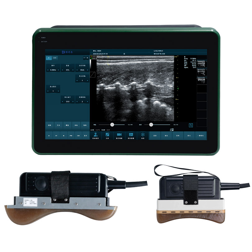
Donkey Pregnancy Detection Using Veterinary Ultrasound
Currently, the most reliable method for early pregnancy detection in donkeys is transrectal ultrasound, which can accurately detect pregnancy in female donkeys around day 16.
Transrectal ultrasound is an important tool for reproductive veterinarians. Toutefois, it cannot replace rectal palpation and should be used in conjunction with it. This technique has its disadvantages, such as the cost of the equipment. Furthermore, transrectal scanning can be difficult between the third and fifth to sixth months of pregnancy because the gravid/pregnant uterus extends into the abdomen, potentially beyond the examiner’s reach. Despite these limitations, this tool is invaluable, particularly for early pregnancy detection, twin management, and fetal vitality assessment.

High-Definition Donkey Ultrasound
Early Pregnancy Detection and Twin Management in Female Donkeys Using Veterinary Ultrasound
Premier, donkey ultrasound can detect an embryonic cyst as early as 9-10 days after ovulation. By day 14-15, the accuracy of cyst detection increases to 99%. One of the most important roles of ultrasound in donkey pregnancy diagnosis is in twin detection and reduction. For initial pregnancy detection, ultrasound examination of the dam between days 13-16 is beneficial. If a synchronous or asynchronous double ovulation is noted, this period is ideal for imaging potential twin bubbles and planning manual reduction of one of the concepts. Between days 16 et 17 after ovulation, increased uterine tone, insufficient uterine contractions, and cessation of embryonic activity lead to fixation of the concepts at the base of one uterine horn. If both twins are fixed to the base of one uterine horn, manual reduction may be more difficult with later detection after days 17-20, known as unilateral fixation. If each conception occupies one uterine horn in bilateral fixation, reduction is easier after bilateral fixation.
Between days 21 et 25, embryos that ultimately develop into fetuses can be detected. If twins are still present, intervention via reduction or transvaginal aspiration is still possible, but the survival rate of the remaining twin is often lower. If twin reduction is performed before this time, the ultrasound can confirm the presence of a single pregnancy. Furthermore, the embryo’s heartbeat can be detected as early as day 22 on some ultrasounds and consistently on day 25 or later on most ultrasounds. If the embryo does not develop normally or the pregnancy is lost, the female donkey can be rebred before the endometrial cup forms. Twin management is discussed in detail elsewhere. It is important to note that two ultrasound examinations should be performed before day 30 of pregnancy to increase the chance of detecting twins.
Ultrasound Testing for Fetal Viability in Female Donkeys
To monitor embryonic and fetal growth, charts can be used to show how the conceptus should appear on ultrasound at specific time points. Familiarity with these images allows a skilled reproductive veterinarian to roughly determine embryonic/fetal age or detect early embryonic loss. Using transrectal ultrasound, practitioners can assess embryonic and fetal viability, evaluate placental health by measuring uterine and placental thickness (CTUP), and determine fetal sex at specific times. Early detection of miscarriage or placentitis allows for more effective treatment and monitoring of the female donkey.
De plus,, transrectal imaging of the genital tubercle between days 55-75 can be used to determine fetal sex. Between days 90 et 150, transrectal ultrasound can be used to determine fetal sex from the genital tubercle. The optimal timing of this later window depends on the location of the pregnant uterus.
Ultrasound Images of Male Donkeys
Although not discussed in detail in this article, transabdominal ultrasound can also be used from day 80 until term to monitor placental health, including heart rate, movement and development, fetal sex, and late twin management.
Overall, it is beneficial to perform multiple scans at specific time points to ensure normal embryonic/fetal growth and viability. If the female donkey has concerns about early embryonic loss, twins, fetal loss, or placentitis, additional ultrasound examinations may be performed based on the veterinarian’s recommendation.
These examinations not only inform owners that their female donkey is pregnant but also allow for improved breeding planning for the following season.