As livestock producers strive for ever-greater efficiency and animal welfare, knowing exactly when a cow is pregnant—and monitoring her gestation—has become essential. Hoy, ultrasonography is a cornerstone of bovine reproductive management. But when did this technology become part of the cattle industry’s toolkit? En este artículo, we’ll explore the evolution of pregnancy diagnosis in cows, track the introduction and adoption of ultrasound, explain how and why we can now detect pregnancy as early as 21 days post-breeding, and consider the global impact of this breakthrough.

Traditional Methods of Pregnancy Diagnosis
Rectal Palpation
Before the advent of ultrasound, the predominant on‑farm technique was rectal palpation. An experienced veterinarian or technician would insert their arm into the rectum to feel the uterus, fetal membranes, y, eventually, the fetus itself.
-
Timing: Accurate detection typically became possible around day 30 of gestation, although skilled practitioners sometimes pushed this boundary a few days earlier.
-
Limitations: Early gestational stages (before 30 days) were nearly impossible to diagnose reliably. There was also a small risk of inducing stress or even abortion if done improperly.
Biochemical Tests
By the late 1970s and 1980s, blood‑ and milk‑based assays detecting pregnancy‑associated glycoproteins (PAGs) and other hormones began to appear.
-
ELISA tests could identify pregnancy by day 28 post‑insemination with high accuracy.
-
These tests offered a non‑invasive alternative but required laboratory processing and often cost more per test than palpation.
The Advent of Veterinary Ultrasonography
Origins in Medical Imaging
Ultrasound technology was first recognized for its potential in imaging in 1939 by Sergei Sokolov, but early equipment lacked the resolution and reliability needed for clinical use. By the 1950s and 1960s, ultrasound had become a mainstay in human obstetrics and cardiology.
First Veterinary Applications
-
Equine Medicine (1980): Gray‑scale “B‑mode” ultrasonic imaging was introduced for examining the reproductive tract of mares around 1980.
-
Bovine Research (1984): Within a few years, veterinarians adapted the same technology to cattle. By 1984, transrectal gray‑scale ultrasound was being used in research settings to characterize ovarian follicle dynamics and detect early pregnancy in heifers and cows.
From Research to On‑Farm Practice
Throughout the late 1980s and 1990s, improvements in ultrasound probe design, portabilidad, and image clarity drove broader adoption:
-
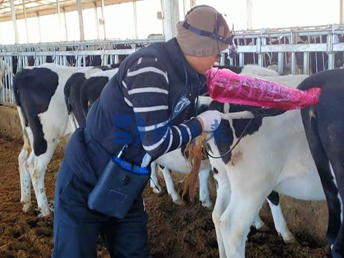
Portable Units: The development of battery‑powered, waterproof scanners made on‑farm ultrasounds practical under field conditions.
-
Operator Training: Extension programs and veterinary schools began offering specialized training in bovine ultrasonography, ensuring a growing pool of skilled users.
By the early 2000s, ultrasound had become a standard tool on progressive dairy and beef operations worldwide.
How Ultrasound Detects Pregnancy in Cows
Principle of B‑Mode Imaging
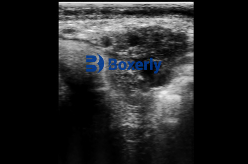
B‑mode (brightness‑mode) ultrasound sends high‑frequency sound waves into the cow’s reproductive tract via a transrectal probe. The echoes reflected from tissues create a two‑dimensional grayscale image:
-
Amniotic Vesicle: Visible as a fluid‑filled sac as early as day 21–26 post‑insemination.
-
Fetal Heartbeat: Detected reliably from day 26 onward.
-
Corpus Luteum & Ovarian Structures: Allow assessment of ovarian function and rule out false positives from cysts.
Early Detection: 21 Days Post‑Breeding
Thanks to enhanced resolution, many practitioners now confirm pregnancy 21 Días after artificial insemination or natural mating. This aligns with physiological markers—around the time when the embryonic vesicle has formed and the fetus begins producing detectable signals.
Key Fact: Artificial insemination or natural mating allows for pregnancy checks as early as 21 Días post‑service.
Advantages of Ultrasound over Other Methods
| Característica | Rectal Palpation | PAG‑Based Tests | Ultrasonography |
|---|---|---|---|
| Earliest Detection | ~Day 30 | ~Day 28 | Day 21–26 |
| Invasiveness | Moderate risk | Non‑invasive | Non‑invasive |
| Speed of Results | Instant | Hours to days | Instant |
| Ability to Assess Fetal Health | No | No | Sí (heartbeat, viability) |
| Repeatability | Limited | Bien | Excellent |
-
Non‑invasive & Safe: No risk of inducing abortion when performed correctly.
-
Real‑time Monitoring: Immediate feedback on pregnancy status, fetal viability, and potential pathologies.
-
Quantitative Data: Measurements of embryonic vesicle size, fetal crown–rump length, and ovarian structures guide management decisions.
Practical Applications on the Farm
Reproductive Management
-
Early Re‑breeding Decisions: Identifying non‑pregnant cows at 21 days means returning them to the breeding pool quickly, reducing days open and improving herd fertility.
-
Embryo Transfer & IVF Programs: Ultrasound guides transcervical embryo transfers and monitors donor cow response to superovulation protocols.
Supervisión del estado
-
Fetal Viability: Detecting a heartbeat by day 26 ensures the embryo is developing normally.
-
Pathology Detection: Ultrasound can reveal uterine infections, luteal cysts, or embryonic loss.
Economic Impact
-
Reduced Feed Costs: Shortening non‑productive days lowers feed expenses per pregnancy.
-
Improved Uniformity: Early detection allows for grouping cows by gestational stage, optimizing nutrition plans.
Global Adoption and Future Directions
Worldwide Uptake
From North America and Europe to South America, Australia, and parts of Asia, ultrasonography is embraced by both large‑scale operations and smaller farms. Veterinary equipment manufacturers offer specialized probes and software tailored for bovine reproduction.
Technological Innovations
-
Ecografía Doppler: Adds blood‑flow assessment to detect placental and embryonic perfusion.
-
3D/4D Imaging: Experimental use for detailed fetal anatomy and volumetric measurements.
-
Artificial Intelligence: Early trials use AI to auto‑measure embryonic structures, reducing operator variability.
Looking Ahead
As probe portability improves and costs decline, even smallholder farmers will gain access. Combined with on‑farm data management and genetic tools, ultrasound will remain pivotal for efficient, welfare‑oriented cattle reproduction.
Conclusión
Ultrasonography revolutionized bovine pregnancy diagnosis when gray‑scale B‑mode imaging was first applied to cows in the mid‑1980s
. What began as a research technique has become the global standard, enabling producers to detect pregnancy as early as 21 days post‑breeding, monitor fetal health in real time, and make data‑driven management decisions. Compared to rectal palpation and biochemical assays, ultrasound offers unmatched speed, safety, and diagnostic depth. As technology continues to advance—with Doppler capabilities, AI‑driven analysis, and 3D imaging on the horizon—the role of ultrasonography in cattle reproduction will only grow.
References
-
“Ultrasound.” Wikipedia, last updated June 2025. https://en.wikipedia.org/wiki/Ultrasound
-
Ginther, O.J., “Gray-scale Ultrasonic Imaging in Equine and Bovine Reproduction.” Theriogenology, 1984. https://pubmed.ncbi.nlm.nih.gov/24274416/
-
Richter, M.P., & Lane, T.J., “The Role of Transrectal Sonography in Pregnancy Diagnosis in Cattle.” Journal of Diagnostic Medical Sonography, 2022. https://www.sdms.org/docs/default-source/jdmspdf/pdf-of-artilce-the-role-of-transrectal.pdf
-
IMV Technologies, “Use of Ultrasound Scanning Technique – Bovine.” 2022. https://www.imv-technologies.com/academy/use-of-ultrasound-scanning-technique-bovine
-
“Cattle Genomics: Pregnancy Diagnosis in Beef Cattle.” University of Tennessee Extension. https://utbeef.tennessee.edu/cattle-genomics-pregnancy-diagnosis-in-beef-cattle/