In the realm of beef cattle production, postpartum uterine health plays a crucial role in ensuring reproductive efficiency and overall herd productivity. One critical physiological process following calving is uterine involution—the return of the uterus to its pre-pregnancy size and function. Timely and effective uterine involution is essential for reducing the interval between calving and successful rebreeding. As modern veterinary technology advances, ultrasonido has emerged as a powerful, non-invasive tool to monitor this involution process. This article explores how veterinary ultrasound technology assists farmers and veterinarians in analyzing the speed of uterine involution postpartum in beef cattle, highlighting both practical applications and scientific insights from global research.

Understanding Uterine Involution in Beef Cattle
Uterine involution refers to the physiological process whereby the uterus gradually shrinks and recovers structurally and functionally after the expulsion of the fetus and placenta. In beef cattle, this process generally spans 30 Para 60 days postpartum but can vary widely based on several factors including breed, nutrición, health status, and management practices.
Effective involution is vital because delayed uterine recovery often leads to reproductive disorders such as endometritis or metritis, which can impair fertility and lengthen the calving interval. The earlier and more accurately involution progress is assessed, the better the management interventions can be tailored to promote reproductive health.
Traditional vs Modern Methods to Monitor Uterine Involution
Historically, uterine involution evaluation relied heavily on manual rectal palpation, which is subjective and limited by the examiner’s experience and the animal’s condition. This method provides rough estimates of uterine size and tone but lacks precision.
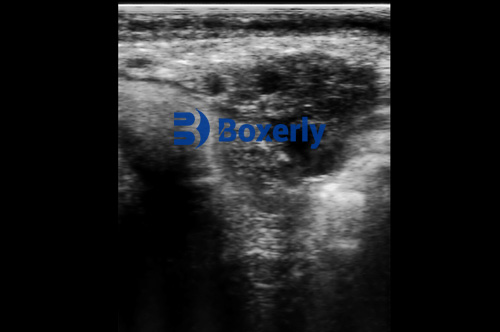
En contraste, veterinary ultrasound technology—particularly B-mode ultrasound—is revolutionizing how uterine involution is assessed. Ultrasound enables visualization of uterine size, endometrial thickness, presence of fluids, and tissue characteristics in real time. This non-invasive imaging technique provides objective and quantifiable data, which improves diagnostic accuracy and aids in early detection of postpartum uterine complications.
Ultrasound Imaging Techniques for Postpartum Uterine Evaluation
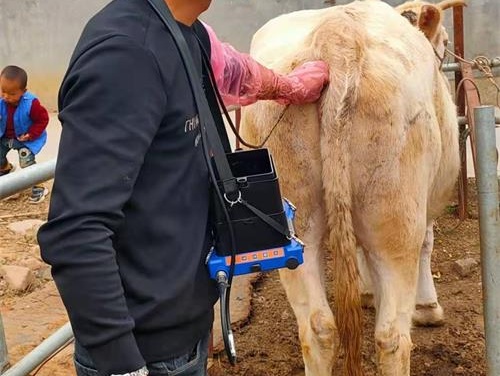
Veterinary ultrasound machines use high-frequency sound waves to generate detailed images of internal structures. For postpartum beef cows, transrectal ultrasound probes are typically used, providing direct access to the reproductive tract.
Key Ultrasonographic Parameters
-
Uterine Horn Diameter: Measurement of each uterine horn’s diameter helps track involution as the uterus gradually shrinks.
-
Endometrial Thickness: Reflects uterine lining regeneration; thickened endometrium may indicate inflammation or delayed recovery.
-
Intrauterine Fluid Volume: Fluid presence is normal shortly after calving but persistence suggests infection or retained placental material.
-
Echo Texture: Changes in uterine wall echogenicity can indicate tissue repair or pathology.
-
Ovarian Status: Monitoring ovarian structures complements uterine evaluation by indicating return to cyclicity.
These parameters collectively provide a comprehensive picture of uterine health and involution speed.
Practical Application on Beef Cattle Farms Worldwide
Veterinarians and livestock managers across the globe have embraced ultrasound for postpartum uterine monitoring due to its several advantages:
-
Early Diagnosis of Postpartum Disorders: Ultrasound detects retained fetal membranes and uterine infections sooner than clinical signs appear, enabling prompt treatment.
-
Guiding Reproductive Management: Understanding involution status informs decisions about the timing of breeding or artificial insemination, minimizing open days.
-
Reducing Economic Losses: By preventing prolonged calving intervals and improving reproductive efficiency, ultrasound monitoring enhances overall profitability.
In the United States, research institutions such as Colorado State University and the University of Missouri have published studies validating ultrasound as an effective tool for postpartum uterine evaluation in beef cows. Similarly, in Australia and New Zealand, where beef production is intensive, ultrasound-assisted reproductive management is integrated into herd health programs to optimize productivity.
Scientific Insights on Factors Affecting Uterine Involution Speed
International studies have identified multiple intrinsic and extrinsic factors influencing involution speed, which ultrasound allows to monitor closely:
Breed Differences
Some beef breeds exhibit faster uterine involution. Por ejemplo, Bos indicus cattle often show slower involution compared to Bos taurus breeds, likely due to genetic and physiological differences. Veterinary ultrasound helps quantify these breed-related variations, assisting selective breeding programs to improve reproductive traits.
Nutritional Status
Adequate nutrition postpartum is critical. Energy deficits or mineral imbalances can delay uterine repair. Ultrasound monitoring combined with nutritional assessment guides feeding management to support faster involution.
Health and Disease
Postpartum infections such as metritis dramatically slow involution. Ultrasound can visualize abnormal uterine contents and wall thickening, facilitating early diagnosis and treatment.
Management Practices
Calving conditions, hygiene, and stress levels impact uterine recovery. In extensive grazing systems, frequent ultrasound checks may be less feasible but remain valuable for high-risk animals. Intensive feedlot systems more commonly use routine ultrasound screening postpartum.
Step-by-Step Ultrasound Protocol for Uterine Involution Assessment
A typical veterinary ultrasound examination of uterine involution postpartum includes:
-
Preparation: Restrain the cow safely; ensure probe and machine are clean.
-
Probe Insertion: Insert transrectal probe gently to access uterus.
-
Image Acquisition: Scan both uterine horns longitudinally and transversely.
-
Medidas: Record uterine horn diameters at consistent anatomical landmarks.
-
Evaluate Fluid and Endometrium: Note presence, volumen, and characteristics of intrauterine fluid; measure endometrial thickness.
-
Ovarian Examination: Document Desarrollo folicular and corpus luteum presence.
-
Repeat Assessments: Conduct serial scans weekly or biweekly to monitor involution dynamics.
Case Example: Ultrasound Monitoring Improving Herd Fertility
In a Canadian beef operation managing 500 Vacas, veterinary ultrasound was implemented postpartum for all cows. Cows showing slower uterine involution or abnormal fluid accumulation received targeted antibiotic and anti-inflammatory treatments. Breeding was postponed until ultrasound confirmed uterine recovery.
After two calving seasons, the herd’s average calving interval decreased by 15 Días, and first-service conception rates increased by 20%. The producer credited ultrasound monitoring as a key factor in enhancing reproductive performance, underscoring ultrasound’s practical value.
Challenges and Limitations of Ultrasound in Uterine Involution
While highly valuable, ultrasound technology is not without limitations:
-
Operator Skill Dependency: Accurate image acquisition and interpretation require training.
-
Animal Handling Stress: Though non-invasive, the procedure needs proper restraint.
-
Equipment Costs: Initial investment and maintenance can be significant, especially for small-scale producers.
-
Field Conditions: In extensive systems, access to electricity or shelter can limit ultrasound use.
Nevertheless, with growing affordability and portable units, these challenges are gradually being overcome.
Future Directions: Innovations in Ultrasound for Beef Cattle Reproduction
Emerging advancements promise to enhance ultrasound’s utility:
-
3D and Doppler Ultrasound: Providing volumetric data and blood flow information for better uterine health assessment.
-
Artificial Intelligence (AI) Integration: AI-driven image analysis could improve diagnostic accuracy and reduce operator variability.
-
Wireless and Handheld Devices: Increasing portability and ease of use on remote farms.
-
Combined Biomarker and Ultrasound Approaches: Integrating ultrasound with molecular diagnostics to refine reproductive health monitoring.
International collaborations among veterinary schools and livestock industries are actively exploring these frontiers.
Conclusión
Veterinary ultrasound technology has become an indispensable tool in analyzing uterine involution speed postpartum in beef cattle. By offering precise, real-time insights into uterine size, tissue condition, and pathological changes, ultrasound supports timely interventions that improve reproductive efficiency and herd profitability.
Global research and practical applications highlight that ultrasound not only enhances reproductive management but also helps reduce economic losses caused by reproductive disorders. As ultrasound devices become more accessible and advanced, their role in postpartum uterine monitoring is expected to expand further.
For beef cattle producers and veterinarians aiming to optimize herd fertility and productivity, integrating veterinary ultrasound into postpartum reproductive protocols is a scientifically validated, practical strategy that delivers measurable benefits.
References
-
Sheldon, I. M., et al. (2009). “Uterine Health and Fertility in Cattle.” Animal Reproduction Science, 110(3-4), 204-214.
https://doi.org/10.1016/j.anireprosci.2008.08.020 -
Peters, Un. R., et al. (2013). “Ultrasonographic Evaluation of Postpartum Uterine Involution in Beef Cattle.” Veterinary Record, 173(20), 503.
https://veterinaryrecord.bmj.com/content/173/20/503 -
Drost, M. (2007). “Ultrasound for Reproductive Management in Beef Cattle.” Theriogenology, 68(3), 456-461.
https://doi.org/10.1016/j.theriogenology.2007.04.029 -
University of Missouri Extension. (2024). “Using Ultrasound to Monitor Uterine Involution in Beef Cows.”
https://extension.missouri.edu/publications/g2107 -
Colorado State University Veterinary Teaching Hospital. (2023). “Postpartum Reproductive Ultrasound in Beef Cows.”
https://csuvth.colostate.edu/reproduction/postpartum-ultrasound/