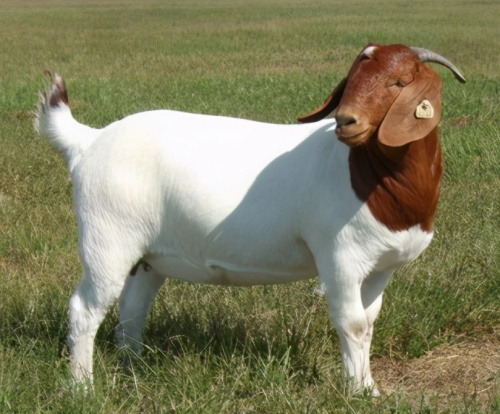
In the modern livestock industry, reproductive efficiency is a cornerstone of profitability, especially for prolific species like Boer goats. Originally bred in South Africa for meat production, Boer goats are now widely raised in many countries due to their rapid growth, good meat quality, and strong reproductive performance. Sin embargo, maximizing reproductive efficiency requires accurate identification of estrus, optimal breeding timing, and monitoring of sexual maturity—tasks that can be challenging without technological support. Among the most powerful tools available, ultrasonography stands out as a non-invasive, precise, and real-time method for monitoring reproductive physiology. En este artículo, we explore how ultrasonography is used to observe the reproductive patterns of Boer goats and how this knowledge helps producers improve breeding outcomes.
Understanding Estrus and Ovulation Timing in Boer Goats
Boer goats are known for being early-maturing and seasonally polyestrous animals. Estrus behavior and ovulation timing vary between young (nulliparous) and adult (multiparous) does. Based on extensive field studies and veterinary observations, most young Boer does ovulate approximately 30 hours after the onset of estrus. En contraste, 75% Para 85% of adult does ovulate closer to 40 hours after estrus onset.
This variation in ovulation timing is critical when determining the optimal breeding window. According to research and practical guidelines, the best time to breed is roughly 5 Para 8 hours before the end of estrus. This corresponds to about 30–35 hours after the first signs of heat for young does, and slightly later for older does. Missing this window can lead to poor conception rates, especially in artificial insemination programs where timing is more sensitive.
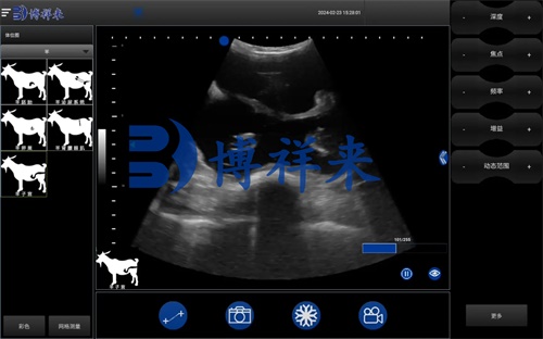
Ultrasound technology plays a vital role in precisely tracking this ovulation window. Real-time B-mode ultrasonography enables veterinarians and producers to visualize the ovaries, detect developing follicles, and determine the exact moment of ovulation. This technology is especially useful when dealing with silent or abnormal estrus cases, where external signs are minimal or misleading.
Monitoring Sexual Maturity and Suitable Breeding Age
While Boer goats may display their first estrus signs (known as the puberty or initial estrus) as early as 6–8 months of age in females and 4 months in males, this does not mean they are ready to breed. En esta etapa, although their reproductive organs start functioning and secondary sexual characteristics are apparent, the animals are not yet physically mature.
For female kids, the initial estrus typically appears when their body weight reaches 40–60% of the adult weight. Males at 4 months may begin to show sexual interest and even ejaculate viable sperm, but both sexes need time for full development of the reproductive tract and associated systems.
International goat farming standards and breeding manuals recommend delaying first mating until female Boer goats reach at least 10 months of age and males are at least 12 meses de edad. Ultrasound examination of the reproductive tract can help confirm that internal organs such as the uterus and ovaries (in females) or testes (in males) have reached maturity, helping avoid problems associated with early breeding such as stunted growth or reproductive complications.
Ultrasonographic Identification of Estrus in Boer Goats
Detecting estrus accurately is the first step in improving reproductive efficiency. For Boer goats, signs of estrus include increased vocalization, tail wagging, mounting behavior, reduced appetite, and swelling or reddening of the vulva. Sin embargo, these signs can sometimes be subtle or absent, especially in animals with nutritional deficiencies or under stressful environmental conditions.
There are three primary methods to detect estrus in Boer does:
-
External Observation
This remains the most commonly used method worldwide. By observing behavioral and physical changes such as tail wagging, restlessness, and vaginal discharge, experienced herders can identify does in heat. Sin embargo, this method depends heavily on the observer’s skill and experience. -
Vaginal Examination
This is a more precise method, especially useful when external signs are unclear. Using a speculum and light source, practitioners can examine the condition of the vaginal mucosa and cervix. During estrus, the vaginal wall appears reddened, moist, and secretes clear mucus. The cervix becomes more relaxed and may open slightly. -
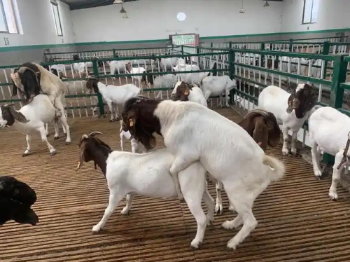
Male Teaser Method
This practical method involves introducing a vasectomized male (teaser) goat into the herd to provoke estrus responses in females. If a female stands still when mounted by the teaser or actively seeks the male’s attention, she is likely in heat. This technique is widely used in both commercial and traditional herding systems around the world.
While these methods are effective, ultrasonography provides an additional layer of precision by allowing direct visualization of ovarian structures, including developing follicles and corpus luteum, which confirms the reproductive stage.
Using Ultrasound to Determine Ovulation and Breeding Time
Ultrasound not only helps detect estrus but also accurately determines ovulation timing—a key to successful timed breeding and artificial insemination. By performing transrectal or transabdominal ultrasound scans at 12- to 24-hour intervals during estrus, operators can observe follicular development and eventual ovulation.
Veterinary researchers and farmers alike have observed that ovulation in Boer does typically occurs around 30–40 hours after the onset of estrus. By monitoring the disappearance of the dominant follicle and formation of the corpus luteum, ultrasound provides definitive evidence that ovulation has occurred. This is essential for scheduling AI procedures or natural mating to maximize fertility rates.
In cases of prolonged estrus or uncertain timing, a second mating or insemination 5–10 hours apart may be recommended. This is especially useful when multiple follicles are present or when animals show intermittent estrus behaviors.
Detecting Abnormal Estrus Patterns with Ultrasonography
Not all Boer goats exhibit typical estrus cycles. Nutritional deficits, stress, illness, or poor management can result in irregular patterns such as:
-
Silent Estrus – When the female ovulates without showing behavioral signs of heat. This is often due to low estrogen levels and is a common reason for missed breeding opportunities.
-
Short Estrus – Where the visible signs of estrus last only a few hours, making detection difficult.
-
Interrupted Estrus – A prolonged, inconsistent display of estrus behavior, often seen in nutritionally stressed animals during late winter or early spring.
Ultrasound is invaluable in these cases, as it can confirm the presence of mature follicles or ovulation, even when behavioral signs are ambiguous.
Global Perspectives: How Other Countries Use Ultrasound in Goat Breeding
Internationally, ultrasonography has become a core tool in small ruminant reproduction programs. In Australia, Canadá, and parts of Europe, B-mode ultrasound is used not only for pregnancy diagnosis but also for follicular mapping, estrus synchronization monitoring, and selection of high-fertility breeding stock.
In large-scale breeding operations, some farms use ultrasound alongside hormonal treatments to synchronize estrus and time AI precisely. This combination has dramatically improved conception rates and reduced the cost of failed breedings. Meanwhile, smallholder farmers in developing countries are increasingly gaining access to portable ultrasound units through veterinary extension services.
Ultrasound is also an important educational tool in veterinary schools and goat breeding training programs worldwide. It allows students and practitioners to visualize physiological processes in real time, strengthening their understanding of reproductive physiology and management strategies.
Conclusión
Ultrasonography is transforming the way Boer goat breeders approach reproduction. By providing real-time, visual insight into ovarian activity, follicle development, estrus detection, and ovulation timing, ultrasound enables more precise and effective reproductive management. Whether on a commercial goat farm in Australia or a smallholder operation in Africa, this technology empowers farmers to make informed decisions, minimize reproductive failures, y mejorar la productividad.
For anyone raising Boer goats, especially those engaged in artificial insemination or selective breeding programs, incorporating ultrasonography into regular reproductive management is no longer optional—it’s essential. With continued technological advancements and broader accessibility, ultrasonography will remain at the heart of efficient and sustainable goat breeding worldwide.
References
-
Smith, B., & Johnson, R. (2020). Ultrasonography in Small Ruminant Reproduction. Journal of Animal Reproduction Science.
-
FAO Small Ruminant Reproductive Management Guide (2023)
-
University of Sydney Veterinary School: Caprine Reproductive Ultrasonography Curriculum (2022)