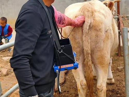
Aspiración con aguja fina (FNA) guiado por Ecografía veterinaria se ha convertido en una piedra angular en la práctica moderna de animales grandes. Se utiliza para tomar muestras de tejidos, como los ganglios linfáticos., hígado, o masas en el ganado, caballos, y pequeños rumiantes, Esta técnica proporciona a los veterinarios un método mínimamente invasivo para recopilar información diagnóstica de forma rápida y eficaz. Aunque la PAAF guiada por ecografía se utiliza ampliamente en las clínicas de pequeños animales, Sus beneficios también se reconocen cada vez más en los entornos ganaderos.
Mejorar en la ecografía por aspiración al afán no solo significa mejores resultados de diagnóstico, sino también una reducción del estrés para los animales, Decisiones más rápidas, y menores costos de tratamiento. Como ganadero o veterinario que trabaja con animales de granja, mejorar su técnica de ultrasonido FNA puede afectar significativamente la salud del rebaño y la eficiencia operativa.
En este artículo, exploraremos formas clave de mejorar sus habilidades en ultrasonido veterinario FNA, guiados por los últimos conocimientos de los organismos veterinarios internacionales y fuentes autorizadas.
Referencias de las autoridades veterinarias:
-
"Aspiración y biopsia con aguja fina guiadas por ecografía": la práctica veterinaria actual, https://todaysveterinarypractice.com/ultrasound-guided-fine-needle-aspiration-and-biopsy/
-
"Ultrasonografía en la práctica ganadera" – Clínicas Veterinarias de América del Norte: Práctica de Animales de Consumo, https://www.sciencedirect.com/journal/veterinary-clinics-of-north-america-food-animal-practice
-
"Ecografía diagnóstica en medicina veterinaria" – Manual veterinario Merck, https://www.merckvetmanual.com
Comprender el propósito de la FNA en animales de granja
La aspiración con aguja fina permite tomar muestras de estructuras internas como los ganglios linfáticos superficiales, abscesos, masas, e incluso ciertos órganos. A diferencia de las biopsias con aguja gruesa, La PAAF es menos invasiva y, a menudo, se puede realizar en el lado de la rampa o en el campo con un equipo de ultrasonido portátil. Cuando se ejecuta correctamente, La PAAF proporciona muestras citológicas que ayudan a detectar infecciones, inflamación, neoplasia, y otras anomalías sin necesidad de intervención quirúrgica.
Equipo y configuración esenciales
Antes de que pueda dominar el procedimiento, Es crucial asegurarse de que está utilizando las herramientas adecuadas:
-
Ecógrafo con transductor lineal o convexo (3.5–8 MHz para animales grandes)
-
Largo, agujas de aspiración de calibre fino (Por lo general, calibre 20-22)
-
5–Jeringas de 10 ml
-
Alcohol o gel para el contacto con la piel
-
Herramientas de recorte para preparar el área
-
Guantes de protección y suministros de campo estériles
Asegúrese de que la máquina de ultrasonido ofrezca funciones de visualización de la aguja, como la dirección del haz o el modo de mejora de la aguja para ayudar a guiar la punta de la aguja con precisión. Portátil, Las unidades alimentadas por baterías son especialmente útiles para operaciones de campo en granjas remotas.
Consideraciones previas al procedimiento
Antes de insertar la aguja, Siga estos pasos::
-
Recorte y limpie el sitio objetivo para reducir la contaminación.
-
Aplique el gel de ultrasonido generosamente.
-
Ajustar la profundidad, ganar, y ajustes de frecuencia para visualizar claramente el objetivo.
-
Practicar la localización de la lesión o masa en los planos longitudinal y transversal.
-
Calmar y sujetar al animal adecuadamente para reducir el movimiento y el estrés.
Incluso un ligero movimiento puede causar errores de muestreo o crear dificultades para mantener la aguja a la vista, Por lo tanto, una buena restricción, ya sea física o química, es esencial.
Técnica ecoguiada: Paso a paso
A continuación, se ofrece una descripción general de cómo realizar una FNA mediante ecografía:
-
Sostenga el transductor en su mano no dominante y visualice el objetivo.
-
Con tu mano dominante, Inserte la aguja en el plano (paralelo a la sonda) o fuera del avión (perpendicular).
-
Mantenga la punta de la aguja dentro del campo de visión: esto es fundamental.
-
Aplique la succión con la jeringa una vez que la aguja esté dentro del objetivo.
-
Mueva la aguja ligeramente hacia adelante y hacia atrás para aspirar las células.
-
Suelte la succión y retire la aguja.
-
Prepare los portaobjetos de muestra inmediatamente para preservar la integridad de la célula.
Consejos para obtener mejores resultados
-
Utilice la trayectoria de la aguja más corta posible para alcanzar el objetivo.
-
Si la visibilidad es escasa, Intente abanicar ligeramente el transductor o ajustar el ángulo.
-
En animales más grandes como ganado vacuno o equino, Los objetivos más profundos pueden requerir sondas de baja frecuencia (p ej., 3.5 MHz).
-
Realizar exploraciones de práctica con regularidad, incluso cuando no se están haciendo FNAs, para mantener las habilidades de imagen.
Interpretación de los resultados de la citología
Lo ideal es que la interpretación citológica la realice un patólogo veterinario capacitado. Sin embargo, Los profesionales de campo aún pueden reconocer ciertas características:
-
Los aspirados purulentos sugieren abscesos o infecciones
-
Las células pequeñas y redondas homogéneas pueden indicar linfoma
-
El material graso puede indicar lipomas o cambios degenerativos
Tenga en cuenta la calidad de la muestra: Los frotis hemorrágicos o mal celulares pueden necesitar una aspiración repetida o un sitio alternativo.
Desafíos comunes y cómo superarlos
-
Mala visibilidad de la aguja:
– Solución: Utilice un ángulo más pronunciado o seleccione el modo de mejora de la aguja en el ultrasonido. -
Animales que no cooperan:
– Solución: Usar una sedación leve o una mejor restricción física. -
Contaminación de la sangre:
– Solución: Evite las estructuras vasculares revisando cuidadosamente el Imagen de ultrasonido.
Educación y Capacitación Continua
Asistir a talleres prácticos y cursos de certificación es una de las mejores maneras de mejorar tus habilidades de FNA. Organizaciones como la Asociación Americana de Practicantes Bovinos (AABP) o la Asociación Británica de Veterinarios de Ganado (BCVA) ofrecer regularmente formación práctica. Adicionalmente, plataformas de aprendizaje en línea como VetMedTeam o VetLexicon ofrecen módulos de aprendizaje virtual.
Conclusión
Aspiración con aguja fina guiada por La ecografía es una herramienta poderosa en la salud del ganado caja de herramientas. Mejorando tu técnica, Perfeccionar la interpretación de la imagen, y mantenerse actualizado a través de la capacitación, Estarás equipado para hacer mejores, Decisiones más rápidas que apoyen tanto el bienestar animal como la productividad de la granja.
Como con cualquier técnica veterinaria, La experiencia y la constancia son la clave. Conviértalo en una parte rutinaria de su enfoque diagnóstico y continúe aprendiendo de cada caso. Sus animales, y su resultado final, se lo agradecerán.