La tecnología de ultrasonido se ha convertido en una herramienta indispensable en la medicina veterinaria moderna. From reproductive management in livestock to diagnosing internal organ abnormalities in companion animals, ultrasound provides veterinarians with a noninvasive and highly informative imaging method. Among the recent innovations, 3D and 4D ultrasound have garnered attention for their enhanced visualization capabilities. But what do these terms truly mean, and how do they differ? More importantly, what role do these technologies play in veterinary diagnostics and farm animal health?
This article explores the technical distinctions between 3D and 4D ultrasound, their applications in veterinary settings, benefits to animal health and farm management, and considerations for integrating them into your practice.
What Is 2D Ultrasound and Why It’s Still Relevant
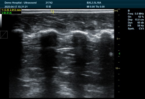
Before delving into 3D and 4D technologies, it’s helpful to understand the foundation: 2D ultrasound. This traditional form of ultrasound uses high-frequency sound waves that travel into the body and bounce back to create a two-dimensional grayscale image of internal organs or tissues. It’s widely used to examine abdominal organs, assess fetal development, guide procedures like fine-needle aspiration, and detect fluid accumulation or abnormalities.
While 2D ultrasound is the workhorse of veterinary imaging due to its speed, Asequibilidad, and real-time feedback, it provides limited spatial perception. The lack of depth or volume perception sometimes makes interpretation challenging—especially when precise anatomical orientation is required.
What Is 3D Ultrasound in Veterinary Medicine?
Three-dimensional (3D) ultrasound builds upon 2D imaging by using multiple sound wave reflections from various angles to construct a volumetric image. Instead of a flat slice, the resulting image has depth and can resemble a photographic reconstruction of the scanned structure.
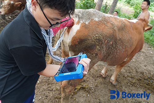
In practical veterinary terms, a 3D ultrasound scan can help visualize anatomical features in a more lifelike manner. This is particularly valuable when evaluating congenital abnormalities in fetuses, tumors with irregular shapes, or reproductive organs in large animals. In livestock applications, 3D ultrasound is increasingly used in advanced reproductive procedures such as ovum pick-up (OPU) and embryo transfer.
The Process Behind 3D Imaging
3D imaging involves either static acquisition (freehand scanning with spatial tracking) or automated scanning using a motorized probe. The captured series of 2D images are processed by specialized software to create a volumetric reconstruction. Once compiled, the image can be rotated, sliced, and analyzed from different perspectives—providing richer information than a single 2D view.
This type of data is especially useful for measuring the volume of organs, assessing tissue architecture, or documenting fetal morphology.
What Is 4D Ultrasound? The Addition of Time
While 3D ultrasound offers static volumetric imaging, 4D ultrasound adds the element of time—essentially creating a real-time video of the 3D image. The “fourth dimension” is motion. With 4D ultrasound, veterinarians and animal breeders can observe movements as they happen within the body.
In fetal monitoring, for example, 4D ultrasound can show a calf or foal moving inside the womb. This dynamic view helps assess vitality, detect abnormalities in real-time, and evaluate functional characteristics such as fetal heartbeat or breathing-like movements. It may also reveal musculoskeletal deformities or behavior patterns that are not obvious in still images.
From a farm management perspective, this technology enables a more precise assessment of gestational progress and viability, helping make timely decisions about breeding cycles and interventions.
Clinical Use Cases of 3D and 4D Ultrasound in Veterinary Practice
-
Reproductive Assessment
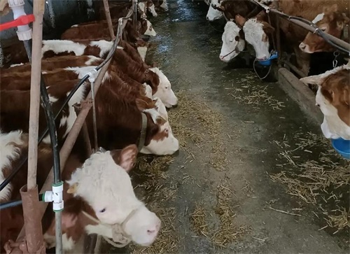
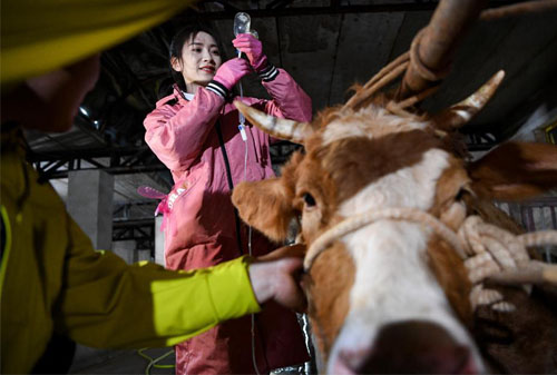
Reproduction is a primary driver of profitability in livestock farming. Early and accurate pregnancy diagnosis, detection of fetal anomalies, and monitoring of ovarian function are critical.
3D ultrasound can visualize the uterus and ovaries in greater detail, facilitating procedures such as follicular aspiration in embryo transfer programs. 4D ultrasound allows observation of embryonic movement, heartbeat, and fetal positioning—offering critical insight into pregnancy health, especially in high-value animals.
-
Detection of Congenital Defects
Just like in human medicine, congenital defects in animal fetuses can lead to complications at birth or loss of valuable livestock. With 3D imaging, veterinarians can identify deformities in the skull, spine, or limbs. Adding 4D motion allows practitioners to see whether muscles and joints are functioning normally, increasing diagnostic confidence. -
Tumor Evaluation and Surgical Planning
In companion animals such as dogs and cats, tumors of the liver, riñones, or mammary glands can be challenging to evaluate using 2D imaging alone. 3D ultrasound provides a better understanding of tumor size, shape, and location, helping plan surgical excisions with greater precision. When observing vascular flow or tumor movement, 4D can offer additional functional insights. -
Guided Procedures
Veterinary interventional procedures—such as biopsies or abscess drainage—benefit from image guidance. 3D ultrasound can provide volume visualization of the target area, and 4D real-time tracking enhances safety and accuracy during needle placement. -
Imágenes cardíacas
In cases where echocardiography is required, 3D and 4D cardiac ultrasound allow for a more comprehensive view of heart valves and chamber dynamics. This is useful in diagnosing valvular insufficiencies or congenital heart defects in young animals.
Operational Benefits of 3D/4D Ultrasound on Farms and Clinics
-
Improved Diagnostic Accuracy
The volumetric perspective of 3D ultrasound reduces the risk of misinterpretation from limited angles, providing more comprehensive anatomical understanding. For time-sensitive reproductive decisions, accuracy means fewer false diagnoses and optimized breeding schedules. -
Enhanced Client Communication
For farm owners or animal caretakers, visualizing a moving fetus in the womb or a clear image of a growth can help them understand the animal’s condition better. This promotes transparency, trust, and quicker decision-making in collaborative care. -
Reduced Need for Repeat Scans
By obtaining more data in one scan, 3D and 4D imaging reduce the need for additional examinations. This is especially beneficial for minimizing stress in large animals or wildlife species where sedation or restraint is a concern. -
Competitive Advantage
Incorporating advanced imaging into your clinic or livestock operation distinguishes you as a forward-thinking provider. This can attract more clients and referrals, especially in regions where access to specialist imaging is limited.
Safety Considerations: Training and Best Practices
While 3D and 4D ultrasound offer many advantages, their usage requires proper training. Improper operation can result in misleading information, which may lead to inappropriate treatments or misdiagnosis.
International regulatory bodies such as the FDA recommend that ultrasound, particularly advanced forms like 3D and 4D, be performed only by trained professionals. This ensures that images are acquired safely, interpreted accurately, and used in the best interest of animal health.
For veterinary professionals new to 3D/4D imaging, investing in comprehensive training programs is essential. Learning how to manipulate, interpret, and apply volumetric data maximizes diagnostic benefits while minimizing the risk of error.
When to Use 3D/4D and When 2D Is Enough
While 3D and 4D ultrasounds offer unparalleled visualization, they are not always necessary. For many routine evaluations—like checking for fluid buildup, assessing bladder stones, or monitoring liver texture—a standard 2D scan provides sufficient information.
Sin embargo, when depth perception, anatomical complexity, or dynamic function is required, 3D/4D ultrasound provides a distinct advantage. It’s not about replacing 2D but expanding your diagnostic toolkit for complex cases.
Practical Tips for Integrating Advanced Ultrasound in Practice
-
Assess Clinical Needs: Evaluate how often you encounter cases where 3D/4D would enhance your diagnostic process.
-
Choose the Right Equipment: Look for systems that offer multi-mode capability—2D, 3D, and 4D in a single unit. Ensure it supports various probes for large and small animals.
-
Budget and ROI: While advanced systems require a higher initial investment, consider the return in terms of improved diagnostics, client satisfaction, and potential service fees.
-
Train Your Staff: Ensure technicians and veterinarians are comfortable acquiring and interpreting images. Many manufacturers or educational institutions offer certified training.
-
Maintain the Equipment: Keep software updated and probes calibrated to ensure optimal image quality and patient safety.
Conclusión: Evolving With Imaging Innovation
As veterinary medicine becomes more advanced, imaging technologies must evolve alongside it. 3D and 4D ultrasounds represent a leap forward in how veterinarians and livestock operators can view, assess, and treat animal health conditions. While not necessary for every case, these tools can be game-changers in reproductive management, surgical planning, and detailed diagnostics.
By embracing these innovations and ensuring proper training, veterinary professionals can provide better care, achieve faster diagnoses, and ultimately improve outcomes for both animals and their caretakers.
Resources
-
American Veterinary Medical Association. “Ultrasound Use in Animal Medicine.”
https://www.avma.org/resources/pet-owners/petcare/ultrasound-use -
DeFrancesco T, Royal K. A survey of point-of-care ultrasound use in veterinary general practice. Education in the Health Professions. 2018;1(2):50.
https://doi.org/10.4103/ehp.ehp_21_18 -
Lyssens A, Lekane M, Gommeren K, et al. Focused cardiac ultrasound to detect pre-capillary pulmonary hypertension. Frontiers in Veterinary Science. 2022; 9.
https://doi.org/10.3389/fvets.2022.830275 -
Feliciano MAR, Uscategui RAR, Maronezi MC, et al. Ultrasonography methods for predicting malignancy in canine mammary tumors. PLoS One. 2017;12(5):e0178143.
https://doi.org/10.1371/journal.pone.0178143 -
Pagani E, Tursi M, Lorenzi C, et al. Ultrasonographic features of adrenal gland lesions in dogs. BMC Veterinary Research. 2016;12(1):267.
https://doi.org/10.1186/s12917-016-0895-1