 BXL
Corpus Luteum Blood Flow in Cattle: How Color Doppler Ultrasound Improves Ovarian Examination
Introducción: Why Is Corpus Luteum Blood Flow Important in Cattle?
Reproductive performance plays a major role in the productivity and profitability of modern cattle farming. For veterinarians, reproductive specialists, and cattle breeding professionals, understanding ovarian function is essential for improving breeding efficiency and managing herd fertility.
The corpus luteum (CL) is a temporary endocrine structure that develops on the ovary after ovulation. It produces progesterone, a hormone that plays a critical role in preparing and maintaining the uterine environment for pregnancy.
One important indicator of corpus luteum development and function is its blood supply.
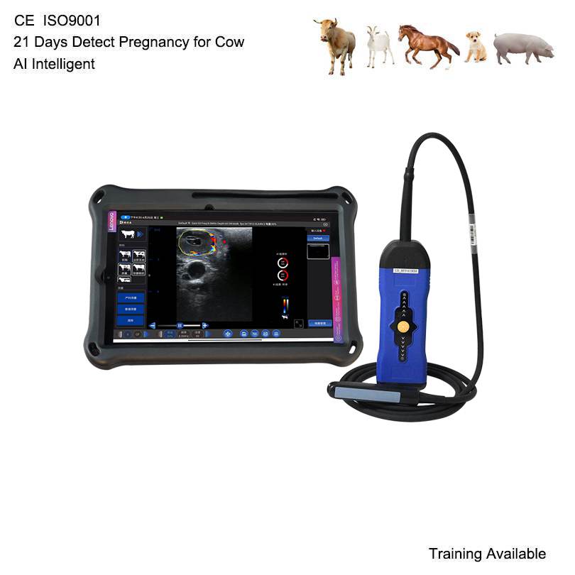
Corpus luteum blood flow is closely associated with luteal development, vascularization, and progesterone production. By using Color Doppler ultrasound, veterinarians can visualize blood flow within and around the corpus luteum, providing additional information beyond conventional grayscale ultrasound imaging.
Hoy, bovine Color Doppler ultrasound is increasingly valuable in reproductive research, ovarian monitoring, and advanced cattle reproductive management.
For veterinarians who need more comprehensive ovarian examination capabilities, a professional veterinary ultrasound scanner with Color Doppler technology can provide significant practical advantages.
1. Understanding Corpus Luteum Development in Cattle
The corpus luteum forms after ovulation when the ruptured ovarian follicle undergoes a series of structural and functional changes.
Following ovulation, the follicular cells develop into luteal cells, while new blood vessels grow into the developing structure.
This process is known as luteal angiogenesis.
The development of an extensive vascular network is essential because the corpus luteum requires an adequate blood supply to deliver nutrients and support progesterone secretion.
The corpus luteum generally undergoes several developmental stages:
Formation: The corpus luteum develops following ovulation.
Growth: Its structure and vascular network become increasingly developed.
Functional maturity: Progesterone production increases as the corpus luteum becomes fully functional.
Regression: If pregnancy does not occur, the corpus luteum normally regresses as part of the estrous cycle.
During these stages, changes in corpus luteum vascularization can provide valuable information about ovarian activity.
This is why corpus luteum blood flow has become an important subject in bovine reproductive ultrasound research.
2. What Is Corpus Luteum Blood Flow?
Corpus luteum blood flow refers to the circulation of blood through the vascular network supplying the luteal tissue.
The corpus luteum is one of the most highly vascularized structures in the ovary. Its blood supply supports the delivery of cholesterol and other essential substances required for steroid hormone production.
In cattle, luteal vascular development is closely related to corpus luteum growth and functional activity.
Evaluating blood flow can help veterinarians and reproductive researchers investigate:
Corpus luteum vascularization.
Luteal development after ovulation.
Changes in ovarian blood perfusion.
Luteal regression during the estrous cycle.
Associations between luteal blood flow and progesterone concentrations.
Ovarian function during reproductive management.
Sin embargo, blood flow should not be interpreted as an independent measurement of progesterone production or pregnancy status. Doppler findings need to be evaluated together with B-mode imaging, reproductive history, and other clinical information.
3. How Does Color Doppler Ultrasound Detect Corpus Luteum Blood Flow?
Color Doppler ultrasound is an advanced imaging technology that provides information about blood movement within tissues.
Unlike conventional B-mode ultrasound, which mainly displays the anatomical structure of the ovary, Color Doppler imaging adds color-coded information representing detected blood flow.
When examining the corpus luteum, the veterinarian positions a transrectal linear probe to identify the ovarian structure.
The scanner can then display color signals corresponding to blood flow within the selected imaging area.
Main applications of Color Doppler imaging include:
3.1 Visualizing Luteal Vascularization
Color Doppler imaging can help identify blood flow signals within and around the corpus luteum.
This allows veterinarians to observe the distribution of vascular signals and assess the vascular appearance of the luteal tissue.
3.2 Monitoring Changes in Blood Perfusion
The vascular pattern of the corpus luteum changes during its development and regression.
Repeated Doppler examinations can help reproductive specialists monitor these changes over time.