For cattle breeders and reproductive veterinarians, understanding the dynamic changes in the bovine ovary is key to optimizing fertility, improving conception rates, and managing herd productivity. In recent years, veterinary ultrasonography has revolutionized the ability to monitor ovarian function in real time, providing detailed insights into follicular waves, ovulation, and corpus luteum (CL) development. In diesem Artikel, we explore how veterinary ultrasound is used to track normal ovarian activity in cows, highlight the patterns of follicular development, and reflect on how this approach is understood and applied by practitioners globally.

Ultrasound and the Ovarian Cycle
Ultraschall für Veterinärmedizin—especially transrectal B-mode ultrasonography—is widely considered the gold standard for evaluating ovarian structures in cattle. It provides a non-invasive, repeatable, and real-time method of visualizing the ovaries, enabling clinicians to identify follicles, detect ovulation, and assess the status of the CL without stress to the animal.
Normal ovarian cycles in cows are characterized by waves of follicular growth and regression, typically involving 2 An 3 distinct waves per estrous cycle. These waves are not just visual patterns—they represent hormonally regulated phases of reproductive readiness, and their detection is essential in synchronization, Züchtung, and embryo transfer programs.
Follicular Dynamics: A Predictable Pattern
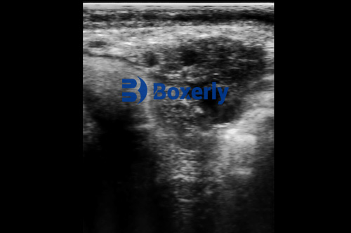
Follicles appear on ultrasound as anechoic (black, fluid-filled) structures that emerge and grow in a clear, rhythmic fashion. Each wave typically begins with a group of small follicles (>5 mm in diameter) that grow concurrently. Darunter, one is selected as the dominant follicle. This dominant follicle then suppresses the development of subordinate follicles through hormonal influence, a phenomenon that is visually observable as the subordinate follicles cease growing while the dominant follicle continues enlarging.
In cows with two-wave cycles, the first wave usually begins shortly after ovulation, with the dominant follicle of that wave becoming atretic (regressing) around Day 11–12 of the cycle. A second follicular wave then begins, giving rise to a new dominant follicle that will ovulate. In three-wave cycles, the second wave also ends in atresia, and it is the third wave’s dominant follicle that eventually ovulates.
Studies in countries like the U.S., Kanada, and Brazil have shown that the timing and expression of these waves can vary slightly with breed, nutrition, and physiological status. Aber, the overarching wave pattern and dominance hierarchy remain consistent and predictable—an essential aspect for reproductive planning.
Ovulation and Its Confirmation via Ultrasound
Detecting ovulation through ultrasound relies on a simple principle: a mature follicle that disappears between successive scans, followed by the appearance of a corpus luteum in the same location, confirms ovulation. Experienced veterinarians often scan cows every 2–4 hours around the expected ovulation window to pinpoint the exact timing.
The corpus luteum, which forms post-ovulation, is a critical structure for maintaining pregnancy. On ultrasound, early CLs appear as irregular, hypoechoic (gray-black) areas within the ovary. By Day 3 post-ovulation, a developing CL can be reliably detected. A mid-cycle CL becomes more structured, with a granular appearance and defined margins. Eventually, the CL regresses if pregnancy does not occur, shrinking in size and becoming less defined.
Veterinarians in Europe and New Zealand often rely on ultrasound for assessing CL function rather than blood-based progesterone testing, especially in field conditions. In fact, studies have shown that the size of the CL measured via ultrasound correlates moderately well with plasma or milk progesterone concentrations (r ≈ 0.68 during functional stages), making it a practical alternative.
Cystic Structures and the Need for Caution
While this article focuses on healthy ovarian cycles, it’s worth mentioning that ultrasonography also plays a role in identifying abnormalities. Follicular cysts—structures that fail to ovulate and continue growing—can be distinguished by their large, thin-walled appearance and lack of accompanying CL. Aber, care must be taken not to confuse a large normal follicle or a cavitated CL with a cyst, especially in the absence of serial observations.
Veterinarians in the U.K. and Australia often emphasize the importance of longitudinal scanning—examining the same cow across several days—to accurately distinguish between normal variation and pathological conditions.
Ultrasound vs. Hormone Testing
Traditionell, progesterone measurement via radioimmunoassay (RIA) or ELISA has been the go-to method for evaluating luteal function. Aber, with the increasing availability of portable ultrasound units, practitioners around the world are shifting toward real-time imaging. Unlike hormone testing, ultrasound provides anatomical information, allowing users to directly observe the presence, size, and structure of the CL and follicles.
This method not only saves time but also reduces reliance on lab infrastructure—an advantage particularly valuable in developing regions or on large farms with time-sensitive breeding schedules. In the U.S., many commercial dairy operations now use ultrasound as a routine herd management tool to guide artificial insemination and detect silent estrus.
Applications Beyond Monitoring
Beyond reproductive cycle tracking, ultrasound has found widespread application in assisted reproductive technologies (KUNST). Zum Beispiel:
-
Embryo Transfer: Pre-transfer scanning ensures the presence of a healthy CL to support implantation.
-
Ovum Pick-Up (OPU): Ultrasound guides follicular aspiration for in vitro fertilization (IVF).
-
Postpartum Uterine Recovery: Scans assess uterine involution and detect infections.
In Latin America and parts of Asia, veterinary ultrasound is often part of a broader strategy to select and manage high-performance cows. By using ovarian status as one of the criteria, producers can more effectively choose cows for breeding, reduce the number of open days, and improve genetic outcomes.
Understanding Follicular Waves in Youthful vs. Mature Cows
Age also influences ovarian dynamics. Young heifers may exhibit more consistent two-wave cycles, while mature, high-yielding milchkühe are more prone to three-wave cycles, possibly due to metabolic differences associated with lactation. Studies in Germany and the Netherlands suggest that energy balance and hormonal metabolism in high-producing cows alter the typical endocrine environment, which may lead to delayed ovulation or persistent follicles.
Deshalb, ultrasound is not just a diagnostic tool but also a window into the complex interaction between physiology and production demands.
The Global Perspective on Ultrasound in Reproductive Management
Across continents, veterinary ultrasound has become a cornerstone of modern cattle reproduction. Its benefits—non-invasiveness, real-time feedback, and visual confirmation—make it a preferred choice for both commercial farms and academic research. In countries like Japan, Italien, and South Africa, national herd improvement programs incorporate routine ultrasonographic checks to improve fertility rates and reproductive efficiency.
Veterinary training institutions worldwide now include extensive modules on ultrasound application, and handheld scanners are becoming more affordable and durable, bringing this technology into the hands of field veterinarians and farmers alike.
Schlussfolgerung
Monitoring normal ovarian activity in cows using veterinary ultrasound offers a reliable and detailed view into the reproductive cycle. By understanding follicular waves, ovulation, and corpus luteum development through real-time imaging, breeders can make better-informed decisions that lead to higher conception rates, shorter calving intervals, and overall improved herd performance.
As global livestock production becomes more sophisticated, ultrasound is no longer a luxury—it’s a necessity. The insights it offers into ovarian dynamics are foundational for reproductive efficiency and sustainable farming. Whether in a research setting or a rural dairy farm, Ultraschall für Veterinärmedizin continues to prove its value, one follicle wave at a time.