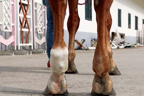
For horse breeders, Tierärzte, and farm managers, ensuring the healthy development of foals is both a responsibility and an investment in the future of the herd. Among the many aspects of neonatal health, proper limb conformation at full term is one of the most critical. Any deviations from normal limb alignment can have lifelong consequences on a horse’s performance, welfare, and economic value. In recent years, advanced ultrasound techniques have become indispensable tools for evaluating foal limb conformation accurately, non-invasively, and at an early stage. This article explores how ultrasonography is transforming fullterm foal limb analysis, why it matters, and how it fits into modern equine reproductive and neonatal management.
Understanding Limb Conformation in Fullterm Foals
Limb conformation refers to the alignment and structural integrity of a horse’s limbs. Ideally, the limbs should be straight, symmetrical, and properly angulated to support the foal’s weight, movement, and future athletic ability. Aber, in many fullterm foals, mild to moderate limb deviations are not uncommon, especially within the first few days after birth.
Common limb deformities include:
-
Angular Limb Deformities (ALDs): Deviations in the angle of the joint when viewed from the front, such as valgus (outward deviation) or varus (inward deviation).
-
Flexural Limb Deformities (FLDs): Abnormal flexion or extension of joints, often related to tendon or ligament contracture.
-
Rotational Deformities: Twisting of the limb along its longitudinal axis.
While many of these issues may self-correct as the foal gains strength and muscle tone, some require early diagnosis and intervention to prevent long-term consequences.
The Growing Role of Advanced Ultrasound in Equine Limb Assessment
Traditionell, radiography (X-rays) has been the primary tool for assessing bone alignment and joint integrity in foals. Aber, X-rays primarily visualize bone and are less effective for soft tissue structures such as tendons, Bänder, cartilage, and joint capsules — all crucial components involved in limb conformation.
This is where advanced ultrasound techniques provide a valuable complement. Ultrasound allows veterinarians to visualize both soft tissue and certain aspects of bone development in real time, offering a more comprehensive evaluation of the foal’s limbs.
Some of the key advantages of using ultrasound for fullterm foal limb analysis include:
-
Non-invasive and radiation-free
-
Real-time dynamic imaging of soft tissues
-
Early detection of subtle abnormalities
-
Ability to monitor growth plates and epiphyseal cartilage
-
Cost-effective and portable for field use
In North America and Europe, veterinary ultrasonography has rapidly gained acceptance in top breeding Bauernhöfe, equine hospitals, and private practices as a standard component of neonatal care.
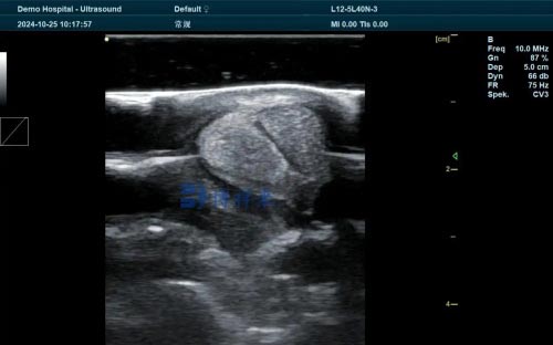
Critical Structures Visualized with Ultrasound
During fullterm foal limb conformation analysis, several critical anatomical structures can be assessed using high-resolution ultrasound:
1. Growth Plates (Physes)
In foals, the growth plates are still open and actively producing new bone. These cartilaginous regions are highly susceptible to developmental abnormalities. Ultrasound can provide clear images of the thickness, uniformity, and integrity of the growth plates, helping to detect early signs of asymmetrical growth that may lead to angular deformities.
2. Articular Cartilage
Joint surfaces are covered with articular cartilage, which cushions and allows smooth joint movement. Ultrasound can detect irregularities, thinning, or damage to this cartilage, which may be involved in joint laxity or instability.
3. Tendons and Ligaments
Flexural deformities often involve the superficial digital flexor tendon (SDFT), deep digital flexor tendon (DDFT), oder suspensory ligaments. High-frequency ultrasound probes allow for detailed visualization of tendon fibers, cross-sectional area, and even early signs of strain or injury.
4. Joint Capsule and Synovial Fluid
Ultrasound can assess joint capsule distension and synovial fluid characteristics, which may indicate inflammation, infection, or developmental joint disease.
5. Subchondral Bone Surface
While ultrasound has limitations in penetrating deep bone structures, it can still reveal surface irregularities of subchondral bone adjacent to articular cartilage, particularly useful in detecting early osteochondral lesions.

Limb Growth Patterns in Fullterm Foals: What Ultrasound Reveals
Foals grow rapidly, especially during the first few months after birth. This growth follows a highly predictable sequence:
-
Nervous system and brain development (mostly completed before birth)
-
Skeletal system growth (including bones and joints)
-
Muscle and tendon maturation
-
Fat deposition (minimal in early foalhood)
At birth, a foal’s limb bones are about 60-70% of their adult length. The remainder of growth occurs through elongation at the growth plates. With serial ultrasound examinations, veterinarians can track whether both sides of the limb are growing symmetrically — an essential factor in preventing angular limb deformities.
Studies from veterinary schools in the United States and Germany have shown that the carpal (knee) and tarsal (hock) regions are particularly vulnerable to growth imbalances that can lead to valgus deformities. Ultrasound helps pinpoint these changes before they become clinically apparent.
Early Diagnosis Leads to Early Intervention
One of the greatest benefits of using advanced ultrasound in fullterm foal limb conformation analysis is the opportunity for early intervention. The earlier deviations are detected, the more treatment options are available, and the better the prognosis.
Common interventions include:
-
Controlled exercise or confinement
-
Corrective hoof trimming and therapeutic shoeing
-
Nutritional adjustments
-
Splinting or bandaging
-
Surgical procedures such as periosteal stripping or transphyseal bridging for severe cases
Ultrasound allows for objective monitoring of treatment progress, guiding veterinarians and farriers in modifying their approach as the foal develops.
Case Example: Ultrasound-Guided Management of Carpal Valgus
On a prominent breeding farm in Kentucky, a Thoroughbred foal was born with a mild carpal valgus deformity (outward deviation of the knee). At three days of age, the attending veterinarian performed a comprehensive ultrasound assessment. The findings showed:
-
Symmetrical growth plate thickness but slightly delayed closure on the medial side.
-
No joint capsule distension or synovial abnormalities.
-
Tendons and ligaments appeared normal.
Based on these ultrasound results, the veterinarian recommended restricted paddock turnout, careful hoof trimming to balance weight-bearing forces, and serial ultrasound exams every two weeks.
By 8 weeks of age, the ultrasound documented complete symmetrical closure of the carpal physes, and the limb deviation resolved without the need for surgery — an outcome that may not have been confidently predicted with radiographs alone.
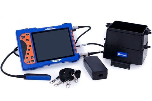
The Technology Behind Advanced Ultrasound
Modern veterinary ultrasound machines offer superior image resolution, Tragbarkeit, and real-time functionality. Devices like the BXL-V50 veterinary ultrasound scanner have become popular among equine practitioners for their versatility:
-
High-frequency linear probes (7–15 MHz) ideal for foal limb imaging.
-
Portable, waterproof design for barn or field use.
-
Real-time Doppler capabilities to assess blood flow in growth plates and soft tissues.
-
Data storage and measurement software for precise growth monitoring over time.
The ability to move between multiple limb sites quickly while capturing high-resolution images allows veterinarians to conduct comprehensive evaluations during a single session, minimizing stress on the foal and improving diagnostic accuracy.
Integrating Ultrasound into a Complete Neonatal Health Program
While ultrasound is an outstanding tool for limb conformation analysis, it functions best as part of a broader neonatal health program. Leading farms in Europe, the U.S., and Australia have developed protocols that combine:
-
Complete physical exams
-
Radiography when appropriate
-
Passive transfer IgG testing
-
Nutritional assessments
-
Daily monitoring of limb alignment and mobility
-
Ultrasound examinations at set intervals (birth, 2 Wochen, 1 month, und 3 Monate)
By combining these tools, breeders maximize the likelihood of identifying and addressing limb conformation issues before they impact the foal’s long-term soundness.
International Perspectives: Growing Adoption of Ultrasound
Veterinary schools and equine specialists worldwide increasingly recognize the value of advanced ultrasound for neonatal limb assessment:
-
In the USA, studies from the University of Florida and Colorado State University highlight ultrasound’s superior sensitivity in detecting subtle growth plate irregularities.
-
In Deutschland, equine orthopedic researchers at the University of Leipzig have pioneered protocols using ultrasound to guide early corrective interventions.
-
In Australien, racing industries rely on early ultrasound monitoring to ensure future performance potential in Thoroughbred yearlings.
As international breeding standards continue to rise, early ultrasonographic evaluation of limb conformation is rapidly becoming a hallmark of responsible, high-quality equine management.
Schlussfolgerung
The health and conformation of a fullterm foal’s limbs set the foundation for its athletic future. Traditional methods like radiography remain useful, but advanced ultrasound techniques have fundamentally elevated the precision, safety, and practicality of neonatal limb assessment.
By visualizing both soft tissue and early bony development, veterinarians can detect subtle deviations that may not yet be visible externally or radiographically. Early detection enables targeted, less invasive interventions that preserve joint integrity, optimize growth, and ultimately enhance the foal’s lifelong soundness and value.
For breeders and veterinarians committed to excellence, investing in ultrasonographic limb analysis represents a forward-thinking approach grounded in both science and compassion — one that is transforming how we safeguard the future stars of the equine world.
References:
-
Reef, V. B. (2021). Equine Diagnostic Ultrasonography (2nd ed.). Wiley Blackwell.
-
Smith, L. J., & Dyson, S. J. (2022). Advances in Ultrasonography of the Equine Neonatal Limb. Equine Veterinary Journal, 54(5), 687-698. https://onlinelibrary.wiley.com/doi/10.1111/evj.13560
-
Rantanen, N. W., & McKinnon, Ein. O. (2017). Equine Reproduction and Ultrasonography. Journal of Equine Veterinary Science, 59, 74-82. https://www.j-evs.com/article/S0737-0806(17)30102-4/fulltext
-
University of Florida Veterinary Medicine (2024). “Using Ultrasound for Early Limb Deformity Detection in Foals.” https://vetmed.ufl.edu/research