Ultrasound is a core diagnostic tool in veterinary medicine, particularly for evaluating soft tissues, Organe, and cardiovascular structures in both large and small animals. In small animals like dogs, echocardiography—a specialized ultrasound technique used to assess the heart—provides invaluable insights into heart morphology and function, as well as inflammatory conditions like myocarditis. As a livestock farmer who also raises companion dogs, I’ve found that understanding the principles and practical techniques of cardiac ultrasound can greatly enhance our ability to detect heart conditions early, treat them effectively, and improve animal welfare.

This article outlines the standard method of performing cardiac ultrasound in dogs and how echocardiography aids in the diagnosis and monitoring of myocarditis, particularly viral-induced types like canine parvoviral myocarditis. It also discusses the veterinary benefits of non-invasive diagnostic imaging and its role in clinical decision-making.
Preparation and Positioning of the Dog for Cardiac Ultrasound
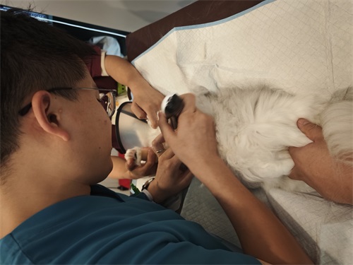
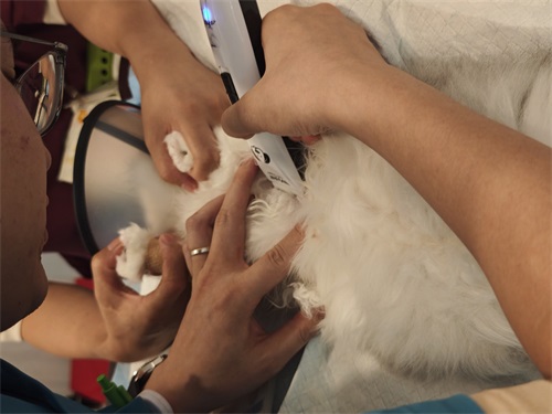
To begin the echocardiographic examination, the dog is typically placed in right lateral recumbency (lying on its right side). Proper restraint and a calm environment are essential to reduce stress-induced cardiac variability. The scanning site is usually between the second and sixth intercostal spaces on the right side of the thorax, most commonly between the fourth and fifth ribs. This area, between the sternum and costal cartilage, is shaved to ensure good contact with the transducer and to minimize signal artifacts.
Ein small animal echocardiographic probe is used with coupling gel to optimize sound transmission. The initial view is obtained using two-dimensional (2D) B-mode ultrasound, which helps locate the heart by identifying precordial pulsations. The probe marker (usually aligned with the operator’s thumb) is oriented so that the cardiac structures appear on the right side of the monitor, with near-field and far-field images clearly delineated. This orientation facilitates the acquisition of a long-axis view of the left ventricle, showing a four-chamber image from the apex of the left ventricle to the base of the right atrium.
This imaging plane is critical for assessing the heart’s structural integrity and observing any abnormal chamber enlargement, valve motion anomalies, or pericardial effusions.
Echocardiography in Diagnosing Myocarditis in Dogs
Myocarditis refers to the inflammation of the heart muscle (myocardium), and in veterinary practice, it can arise from various causes, einschließlich viral infections, electrolyte imbalances, and systemic illnesses. In dogs, canine parvovirus (CPV) is a significant viral agent that can target the myocardium, especially in puppies, leading to sudden cardiac failure or chronic cardiac dysfunction if not detected early.
Experimental Model: Hyperkalemia-Induced Cardiac Dysfunction
A typical model to study cardiac function under stress is the induction of acute hyperkalemia via intravenous administration of potassium chloride (KCl). This mimics severe electrolyte imbalance and helps evaluate how elevated potassium levels affect cardiac structure and performance.
Using 2D echocardiography and pulsed-wave Doppler, we can monitor changes in systolic and diastolic function. Zum Beispiel, hyperkalemia may cause reduced contractility and arrhythmias, which are evident on the echocardiogram. In clinical settings, this helps veterinarians recognize dangerous metabolic conditions before they escalate into heart failure.
Canine Parvoviral Myocarditis
In cases of CPV-induced myocarditis, echocardiography reveals myocardial thinning, chamber dilation, and reduced ejection fraction, all signs of compromised heart function. The virus aggressively replicates within myocardial cells, leading to inflammation, necrosis, and fibrosis. These structural changes are detectable on echocardiography even in the early stages, making it a vital tool for diagnosis.
Unlike some imaging modalities, echocardiography is non-invasive and repeatable, allowing for regular monitoring without causing distress to the patient. This is particularly important for young, immunocompromised animals with CPV, who may not tolerate more invasive procedures.
Techniques and Image Interpretation
Veterinarians performing echocardiography must be skilled in identifying both normal cardiac anatomy and pathological changes. Several key techniques are involved:
-
M-mode Echocardiography
-
This method captures motion over time and is ideal for evaluating the movement of ventricular walls and heart valves.
-
M-mode is used for measuring parameters like left ventricular internal dimension, fractional shortening, und wall thickness during systole and diastole.
-
-
-
Pulsed-wave and color Doppler modalities assess blood flow velocity and direction, which are crucial for detecting regurgitation, stenosis, oder intracardiac shunts.
-
In myocarditis, turbulent flow or altered filling patterns can be visualized.
-
-
Short-axis and Long-axis Views
-
Das short-axis view gives cross-sectional images of the ventricles and allows measurement of wall motion and chamber diameter.
-
Das long-axis view helps visualize the four-chamber structure and assess overall cardiac geometry.
-
By combining these modalities, the examiner can detect even subtle signs of cardiac inflammation, chamber dilation, or reduced myocardial contractility—hallmarks of conditions like viral myocarditis.
Clinical Significance and Veterinary Applications
The integration of echocardiography into small animal practice has several advantages:
-
Early Detection: Subclinical heart changes, especially from viral infections or electrolyte disturbances, can be spotted early.
-
Non-Invasive Monitoring: Echocardiography is gentle enough for fragile patients and can be repeated for treatment monitoring.
-
Guiding Treatment: Based on echocardiographic findings, veterinarians can adjust fluid therapy, prescribe cardioprotective drugs, or recommend hospitalization in severe cases.
-
Disease Research: In academic and clinical research, echocardiography provides visual evidence of disease progression and response to treatment, contributing to better understanding of pathophysiology.
Limitations and Operator Dependence
While echocardiography is a powerful diagnostic tool, it is operator-dependent. Accurate interpretation requires:
-
Deep knowledge of normal canine cardiac anatomy
-
Experience in probe positioning and view optimization
-
Skill in differentiating between artifact and pathology
Zusätzlich, acoustic window limitations due to the lungs or chest wall can hinder image clarity. Yet, with correct positioning and probe manipulation, these obstacles are generally manageable in small animals.
Schlussfolgerung
Cardiac ultrasonography, especially echocardiography, plays a critical role in evaluating heart conditions in dogs. From routine cardiac assessments to diagnosing complex conditions like myocarditis caused by canine parvovirus oder hyperkalemia-induced dysfunction, ultrasound offers a non-invasive, real-time view into the heart’s structure and performance.
As a livestock and dog owner, learning to understand and utilize these imaging techniques enhances the quality of care I can provide and allows for timely veterinary intervention, ultimately improving survival and wellbeing in affected animals. With continuing advancements in veterinary imaging, echocardiography will only grow more accurate, accessible, and impactful in both clinical and field settings.