As a livestock farmer or veterinary professional working with goats, understanding the ovarian follicular dynamics during the estrous cycle is crucial to improving reproductive efficiency and herd productivity. Among various reproductive monitoring techniques, الموجات فوق الصوتية البيطرية technology has emerged as a non-invasive, مصدق, and practical method for real-time visualization of ovarian activity, follicular development, and overall reproductive status in goats.
This article explores how ultrasound technology is applied to monitor ovarian follicular dynamics throughout the goat estrous cycle. It also discusses the underlying biological processes, the benefits of ultrasound monitoring, practical considerations, and how foreign experts have integrated this technology into goat reproductive management.
Understanding Ovarian Follicular Dynamics in Goats
The estrous cycle in goats typically lasts about 18 ل 24 أيام, comprising four stages: proestrus, شبق, metestrus, and diestrus. The cycle is regulated by complex hormonal interactions driving the growth, selection, maturation, and regression of ovarian follicles. Follicles undergo dynamic changes, with several waves of follicular growth occurring each cycle, but usually only one dominant follicle reaches ovulation.
Follicular waves consist of cohorts of follicles that begin growing simultaneously. Among these, one follicle becomes dominant while others regress. The dominant follicle produces estradiol, triggering estrus behavior and ovulation. Monitoring these changes provides insight into the timing of ovulation, fertility status, and potential reproductive disorders.
Role of Veterinary Ultrasound in Monitoring Follicular Dynamics
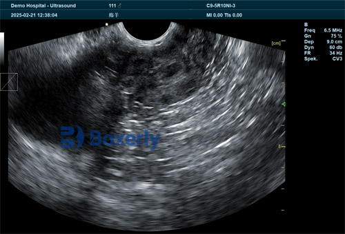
Ultrasound imaging, especially B-mode ultrasound, is a valuable tool to observe ovarian structures in live goats without harm or stress. A transrectal or transabdominal probe can be used to visualize the ovaries and track follicles’ size, رقم, and development stage over the cycle.
How Ultrasound Tracks Follicular Growth
-
Visualization of follicles: Follicles appear as anechoic (dark) circular structures within the ovary on ultrasound images.
-
Measuring follicle diameter: Follicle size measurement helps distinguish dominant follicles from subordinate ones. Dominant follicles typically reach sizes of 5-8 mm or more before ovulation.
-
Follicular wave identification: By scanning every 1-2 أيام, one can observe new follicular waves, the growth and regression phases, and pinpoint the ovulation time.
-
Correlating with estrous behavior: Combining ultrasound data with observed estrus signs improves accuracy in detecting the optimal insemination window.
International Perspective on Ultrasound Use in Goat Reproduction
Globally, veterinary researchers and breeders have increasingly adopted ultrasound as a standard technique to study follicular dynamics in goats, appreciating its accuracy and repeatability.
For example, studies from Spain and Italy highlight ultrasound’s ability to precisely characterize follicular waves and optimize artificial insemination timing. In the United States, ultrasound monitoring supports advanced reproductive technologies like superovulation and embryo transfer, where timing follicular status is vital.
Veterinary colleges across Europe incorporate ultrasound training focused on small ruminants, including goats, emphasizing practical skills in estrous cycle monitoring and fertility management.
Benefits of Veterinary Ultrasound in Managing Goat Reproduction
-
Non-invasive and safe: Unlike hormonal assays or laparoscopic examinations, ultrasound is non-invasive, does not require sedation, and can be repeated frequently without risk to the animal.
-
Real-time visualization: Allows veterinarians and farmers to see follicular growth and regression live, enabling timely decisions.
-
Improved breeding management: Accurate detection of estrus and ovulation enhances the success rates of natural mating and artificial insemination.
-
Diagnosis of reproductive disorders: Ultrasound can detect ovarian cysts, anovulatory follicles, and other abnormalities impacting fertility.
-
Cost-effectiveness: Early identification of reproductive issues helps reduce wasted breeding attempts and improves overall herd productivity.
-
Supports advanced reproductive techniques: Enables effective superovulation protocols, embryo flushing, and transfer.
Practical Application of Ultrasound in Goat Estrous Monitoring
Veterinary professionals or trained farmers perform ultrasound scanning on goats usually every 1-3 days during the estrous cycle to track ovarian follicles. Common steps include:
-
Preparation: Restrain the goat gently to minimize stress. For transrectal scanning, ensure proper probe lubrication and hygiene.
-
Imaging: Place the probe rectally or on the lower abdomen, depending on the goat’s size and ultrasound equipment.
-
Recording follicle size and number: Document follicles’ diameters and observe changes over days.
-
Correlating data: Combine ultrasound findings with hormonal data (if available) and behavioral observations to predict ovulation accurately.
Challenges and Limitations
While ultrasound is an excellent tool, some challenges exist:
-
Skill requirement: Accurate interpretation demands training and experience.
-
Equipment cost: Quality ultrasound machines may represent a significant initial investment.
-
Goat anatomy: Smaller size and different anatomical features can make imaging more difficult than in larger animals.
-
Frequency of scanning: Requires regular scanning to observe follicular waves effectively, which may be labor-intensive.
Despite these, the benefits outweigh challenges, especially when used as part of an integrated reproductive management program.
Case Study: Ultrasound Monitoring Impact in a Commercial Goat Farm
At a commercial dairy goat farm in France, ultrasound monitoring was implemented to improve kidding rates. Prior to ultrasound use, inseminations were timed based on estrus signs alone, resulting in suboptimal conception rates around 50-60%. By adopting ultrasound scanning to identify dominant follicles and optimal insemination timing, conception rates improved to over 75%.
Additionally, early detection of ovarian cysts and other disorders through ultrasound allowed timely veterinary interventions, reducing reproductive wastage. The farm also used ultrasound data to design better hormonal synchronization protocols for breeding, further enhancing productivity.
The Future of Veterinary Ultrasound in Goat Reproductive Management
Emerging advancements in ultrasound technology promise even greater capabilities:
-
Portable and wireless devices: Increasing field usability and convenience.
-
3D and Doppler ultrasound: Offering improved visualization of blood flow in follicles and ovarian الانسجه, giving deeper insights into follicle health and function.
-
Integration with AI: Automated follicle detection and measurement to assist practitioners with faster, more accurate assessments.
-
Remote diagnostics: Enabling experts to guide field practitioners via telemedicine platforms.
These innovations will likely make ultrasound monitoring more accessible, precise, and central to small ruminant reproductive management globally.
استنتاج
Veterinary ultrasound technology is a game-changer in monitoring ovarian follicular dynamics during the goat estrous cycle. By enabling detailed, real-time visualization of follicular growth and regression, ultrasound supports accurate estrus detection, optimizes breeding timing, and facilitates early diagnosis of reproductive disorders.
Across the world, veterinarians and farmers recognize ultrasound as an essential tool for enhancing goat reproductive efficiency and herd profitability. While requiring some expertise and investment, the return on improved fertility and reduced reproductive losses makes ultrasound monitoring invaluable.
As technology advances, ultrasound applications in goat reproduction will become even more powerful, supporting sustainable and efficient goat farming worldwide.
References
-
Morató, R., et al. (2014). “Ultrasound Monitoring of Follicular Dynamics in Goats.” Small Ruminant Research, 120(2-3), 129-137. https://doi.org/10.1016/j.smallrumres.2014.06.001
-
Abecia, J.A., et al. (2017). “Applications of Ultrasonography in Small Ruminant Reproduction.” Theriogenology, 94, 112-119. https://doi.org/10.1016/j.theriogenology.2017.01.036
-
Delgado-Pertíñez, M., et al. (2020). “Follicular Dynamics in Goats and Its Application to Assisted Reproduction.” Reproduction in Domestic Animals, 55(1), 38-47. https://doi.org/10.1111/rda.13515
-
Ginther, O.J. (2007). Ultrasound Imaging and Animal Reproduction: Basic Principles and Clinical Applications. Cross Publishing.