Maintaining the health of the gastrointestinal (GI) tract in livestock is essential to ensure optimal growth, productivity, and economic return. For both pigs and cattle, GI tract disorders are among the most common and economically significant health challenges faced by farmers worldwide. From parasitic infestations to digestive blockages and ulcers, such conditions can impact feed conversion efficiency, weight gain, and overall herd performance. While physical exams and blood tests remain important, ultrasound technology—specifically veterinary B-mode ultrasonography—has emerged as a crucial, non-invasive diagnostic tool to detect and monitor GI tract abnormalities in real-time.
في هذه المقالة, we explore how ultrasound is applied in evaluating GI tract issues in pigs and cattle, what indicators to look for, how foreign veterinarians interpret findings, and why the technique is gaining popularity in livestock operations globally.
Understanding GI Tract Issues in Livestock
Before diving into the role of ultrasound, it’s important to understand the types of GI problems commonly observed in pigs and cattle:
In Cattle
-
Abomasal displacement (Left or Right Displaced Abomasum – LDA/RDA)
-
Rumen bloat و impaction
-
Peritonitis
-
Intestinal volvulus or torsion
-
Foreign body syndrome (hardware disease)
In Pigs
-
Ileitis (caused by Lawsonia intracellularis)
-
Gastric ulcers
-
Enteritis or colitis
-
Intestinal obstruction or intussusception
-
Peritonitis due to bacterial infection or trauma
Many of these conditions are difficult to diagnose externally, particularly in early stages, which is where ultrasonography offers its greatest benefit.
Why Ultrasound?
Ultrasound is an ideal tool for examining the GI tract of pigs and cattle for several key reasons:
-
Non-invasive and stress-free: No need for surgical procedures or sedation.
-
Real-time imaging: GI motility, fluid accumulation, and wall thickness can be directly visualized.
-
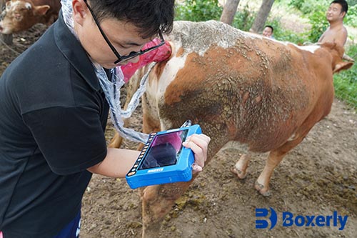
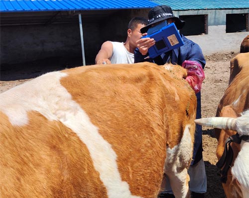
Portable and field-friendly: Devices like the BXL-V50 allow farmers and vets to perform exams on-site.
-
Objective and repeatable: Multiple assessments over time provide data for disease progression or treatment response.
According to international studies and field experience, ultrasound has become an indispensable method for diagnosing internal disorders without resorting to invasive measures.
Ultrasound Technique: How to Scan the GI Tract
In ماشية, ultrasound examinations are typically done from the left or right flanks, depending on the suspected condition. A 3.5–5 MHz convex or linear transducer is used to scan through the abdominal wall.
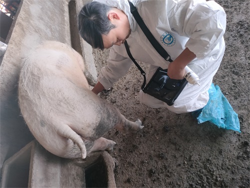
In الخنازير, a 5–7.5 MHz linear transducer is ideal due to their relatively thinner body wall and smaller size. Scanning is usually done in the ventral abdomen or right flank.
Steps for effective scanning:
-
Clip and clean the examination site.
-
Apply ultrasound gel liberally for better image conduction.
-
Begin with longitudinal and then transverse sweeps.
-
Observe GI motility, presence of gas/fluid, wall structure, and neighboring organs.
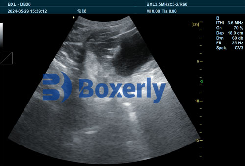
Ultrasound images reveal important clues: thickened intestinal walls may indicate inflammation, while distended loops can suggest obstruction. Fluid pockets or free abdominal gas may suggest rupture or severe infection.
Case Applications: Real-world Examples
1. Detecting Abomasal Displacement in Cattle
Abomasal displacement is one of the most common causes of digestive issues in dairy cows, particularly post-partum. Ultrasound can detect a fluid-filled abomasum with hyperechoic gas interfaces, typically on the left flank for LDA and right for RDA. International guidelines recommend ultrasonography as a first-line diagnostic method, especially for early detection.
2. Diagnosing Ileitis and Ulcers in Pigs
Veterinarians in the EU and North America often rely on ultrasound to detect ileitis, which presents as thickened ileum walls and slowed peristalsis. Gastric ulcers may appear as hypoechoic zones within the stomach. Though sometimes subtle, trained eyes can catch signs early—before symptoms like black stools or sudden death occur.
3. Rumen Disorders and Foreign Body Syndrome
The ultrasound can help distinguish between rumen bloat, foreign body penetration, و reticuloperitonitis. The latter often shows echogenic fibrin and fluid pockets between the reticulum and diaphragm. In some cases, metallic foreign bodies can be indirectly inferred by abnormal movement or abscess formations.
What Foreign Veterinarians Look For
According to the Veterinary Clinics of North America: Food Animal Practice and practical field reports from the UK, كندا, and Australia, trained professionals rely on the following sonographic indicators:
| Indicator | Interpretation |
|---|---|
| Intestinal Wall Thickness | Inflammation or infection |
| Absence of Motility | Paralysis or obstruction |
| Free Fluid in Peritoneum | Peritonitis, rupture, or hemorrhage |
| Dilated GI Loops | Obstruction or ileus |
| Echogenic Material | Feed buildup, blood, or pus |
| Hyperechoic Gas Shadows | Bloat, perforation, or fermentation |
In Denmark, routine GI tract screening in high-value breeding sows is becoming more common, especially with increasing concerns over antimicrobial resistance—prompt diagnosis avoids overuse of antibiotics.
Advantages Over Other Diagnostic Methods
| Method | Invasiveness | Accuracy | Cost | Field Usability |
|---|---|---|---|---|
| Blood Tests | Low | Medium | Low | High |
| Radiography | Medium | High | High | Low |
| Exploratory Surgery | High | Very High | Very High | Very Low |
| Ultrasound | Low | High | Medium | Very High |
Ultrasound offers the perfect balance: accurate enough for clinical decisions, gentle enough for repeated use, and practical for field conditions.
Improving Outcomes on the Farm
On our farm, the adoption of ultrasound for GI tract monitoring has transformed how we handle digestive problems. In pigs, we’ve been able to catch early signs of ileitis and treat with minimal antibiotic use, preserving gut integrity. In cows, early detection of abomasal displacement allows for non-surgical repositioning before the cow becomes critically ill.
By performing routine ultrasound checks on animals with reduced appetite, changes in dung, or abdominal bloating, we’ve significantly reduced emergency interventions and improved animal welfare. Other producers globally, especially in Australia and Germany, are integrating similar routines with excellent results.
Training and Interpretation
One challenge remains: proper interpretation of ultrasound images. That’s why global institutions like the University of Calgary Veterinary Medicine و Royal Veterinary College (UK) now include GI-focused ultrasound modules in their large animal curricula. Devices like the BXL-V50, which feature real-time imaging, simplified controls, and high-contrast displays, help bridge the experience gap for on-site users.
Some basic training enables farmers and technicians to identify obvious abnormalities, while complex findings should still be interpreted by a licensed veterinarian.
Looking Ahead: Preventative and Precision Livestock Management
The future of livestock care is shifting toward preventive management. By routinely scanning the GI tract of young and adult pigs and cattle, we can catch abnormalities before they lead to severe illness or economic loss.
When combined with nutrition planning, stress management, and herd monitoring, ultrasound becomes part of a precision livestock strategy—tailoring interventions based on what’s actually happening inside the animal’s body, not just what’s visible on the outside.
استنتاج
Ultrasound has emerged as a vital, practical tool in diagnosing and managing gastrointestinal issues in pigs and cattle. By providing real-time, non-invasive insight into intestinal and abdominal health, this technique helps producers make faster, more accurate decisions—improving outcomes, animal welfare, and economic efficiency.
Whether detecting displacement, إلتهاب, or obstruction, ultrasound enables early, accurate interventions. As more livestock owners around the world adopt this technology, supported by portable, field-ready equipment like the BXL-V50, the future of GI health management in pigs and cattle looks brighter than ever.
Reference Sources:
-
Braun, U. (2018). Ultrasonography of the gastrointestinal tract in cattle. Veterinary Clinics of North America: Food Animal Practice, 34(1), 69–89. https://doi.org/10.1016/j.cvfa.2017.10.002
-
Whitaker, D. A., & Smith, E. (2021). Veterinary Ultrasonography in Food-Producing Animals. Journal of Veterinary Imaging.
-
Rademacher, C., & Davies, P. (2022). Real-time ultrasound for on-farm swine diagnostics. Swine Health and Production Journal. https://www.aasv.org/shap/issues/v30n4/v30n4rademacher.html
-
University of Calgary Faculty of Veterinary Medicine. (2024). “GI tract imaging in livestock.” https://vet.ucalgary.ca/gi-ultrasound-cattle