Veterinary ultrasonography has become an essential tool in small animal cardiology, particularly in evaluating the structure and function of the canine heart. في السنوات الأخيرة, the development of portable, high-resolution ultrasound devices has enabled veterinarians to perform fast, accurate, and non-invasive cardiac assessments, both in clinics and on-site. Among companion animals, dogs are especially susceptible to various congenital and acquired heart diseases. Detecting and monitoring these conditions early through ultrasound has transformed the approach to canine cardiac care, especially in Western countries where veterinary imaging is a standard practice.
في هذه المقالة, we explore how ultrasound technology is applied in the comprehensive evaluation of a dog’s heart, focusing on the left ventricle, interventricular septum, and right heart chambers. Drawing on current global practices, we also highlight how echocardiographic findings support early diagnosis and treatment of heart failure, valve disorders, and other cardiovascular conditions.
Understanding Canine Cardiac Ultrasound Imaging
Ultrasound imaging—also called echocardiography when used in heart diagnostics—is a real-time, non-invasive method that uses high-frequency sound waves to create visual representations of the heart’s structure and motion. In veterinary cardiology, two main ultrasound modes are commonly used:
-
2D Ultrasound (B-mode): Produces a real-time cross-sectional image of the heart and its structures.
-
M-mode (Motion mode): Displays motion of heart walls and valves along a single ultrasound line over time, ideal for measuring heart wall movement and chamber dimensions during the cardiac cycle.
These imaging modes allow clinicians to measure chamber sizes, observe valve function, assess wall thickness, and evaluate blood flow when paired with Doppler modalities.
Left Ventricle and Interventricular Septum Evaluation
One of the most critical areas in cardiac ultrasound examination is the left ventricle (LV), responsible for pumping oxygen-rich blood to the body. In a healthy dog, the 2D ultrasound image of the LV wall appears as a relatively uniform, low-echogenic structure (a region with fewer echoes). The outer and inner boundaries of the LV are clearly delineated by strong echogenic lines, indicating the edges of the myocardial wall.
M-mode ultrasound reveals a rhythmic wave pattern of the LV wall as it contracts and relaxes. The LV cavity, situated between the LV wall and the interventricular septum, appears as an anechoic (dark, echo-free) oval shape. This region’s size and motion change throughout the cardiac cycle, with M-mode illustrating the alternating phases of diastole (relaxation) and systole (contraction).
ال interventricular septum (IVS) is the muscular wall dividing the left and right ventricles. On 2D ultrasound, it appears as a narrow, low-echogenic band with clearly marked boundaries. M-mode demonstrates its synchronized, yet opposite, movement relative to the left ventricular wall—creating a peak-and-trough pattern with each heartbeat. Any abnormal motion or thickening of the IVS can be indicative of hypertrophic cardiomyopathy or left ventricular outflow tract obstruction.
Right Ventricle and Its Diagnostic Challenges
The right ventricle (RV) lies between the IVS and the right ventricular free wall. On 2D ultrasound, the RV chamber usually occupies about one-third to one-half of the total ventricular view. Due to its anatomical position and proximity to the ultrasound transducer, imaging the RV wall is often more challenging. The image resolution may be reduced in the near field, and fine details are harder to capture compared to the LV.
لكن, with M-mode, the RV wall’s motion can still be visualized, though the amplitude of its motion tends to be smaller. Identifying right-sided dilatation or abnormal wall motion is vital for diagnosing conditions like right-sided heart failure or pulmonary hypertension.
Detecting Cardiac Disease in Dogs with Ultrasound
Ultrasound is not only used for structural evaluation but also plays a key role in diagnosing canine cardiac diseases. For example:
-
Hypertrophic Changes: Some dogs present with thickened interventricular septa and LV walls, along with a reduced LV cavity. This indicates concentric hypertrophy, often seen in pressure overload conditions like aortic stenosis.
-
Right Heart Dilatation: Dilated right ventricles and atria often suggest right-sided heart failure, which may be secondary to tricuspid valve disease or pulmonary hypertension.
-
Mitral Valve Abnormalities: In many dogs, the anterior leaflet of the mitral valve moves abnormally, touching the septal endocardium during systole. This phenomenon, known as systolic anterior motion (SAM) of the mitral valve, can cause left ventricular outflow tract obstruction (LVOTO). It leads to turbulent blood flow and contributes to cardiac dysfunction.
-
Aortic Valve Disease: In some cases, ultrasound reveals abnormal closure of the aortic valve during systole, suggesting structural abnormalities or degenerative valve disease.
Doppler Ultrasound in Advanced Canine Cardiology
Adding Doppler ultrasound to the examination enables the evaluation of blood flow velocity and direction. This is essential for identifying regurgitant flows (backward blood flow), turbulent flows, and pressure gradients across valves.
Continuous wave Doppler and pulsed Doppler modalities are especially useful in dogs showing signs of heart failure. For instance, in mitral valve insufficiency, the Doppler trace at the mitral valve may show:
-
Slowed inflow velocity
-
Disrupted E and A wave patterns (representing early and late diastolic filling)
-
Evidence of increased left atrial pressure
In such cases, Doppler measurements help confirm diastolic dysfunction and guide treatment strategies.
Global Perspectives: How Western Vets Approach Canine Cardiac Ultrasound
In countries like the United States, كندا, and many parts of Europe, canine cardiac ultrasound has become a routine diagnostic tool, especially in:
-
Aging dogs with breed predispositions to heart disease (e.g., Cavalier King Charles Spaniels, Dobermans)
-
Pre-surgical assessments to evaluate anesthetic risk
-
Dogs with clinical signs such as coughing, exercise intolerance, or syncope
Western veterinary practices often follow standardized protocols, such as those from the American College of Veterinary Internal Medicine (ACVIM), which recommend annual screening for dogs at risk and longitudinal monitoring for dogs already diagnosed with cardiac conditions.
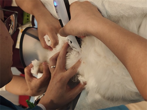
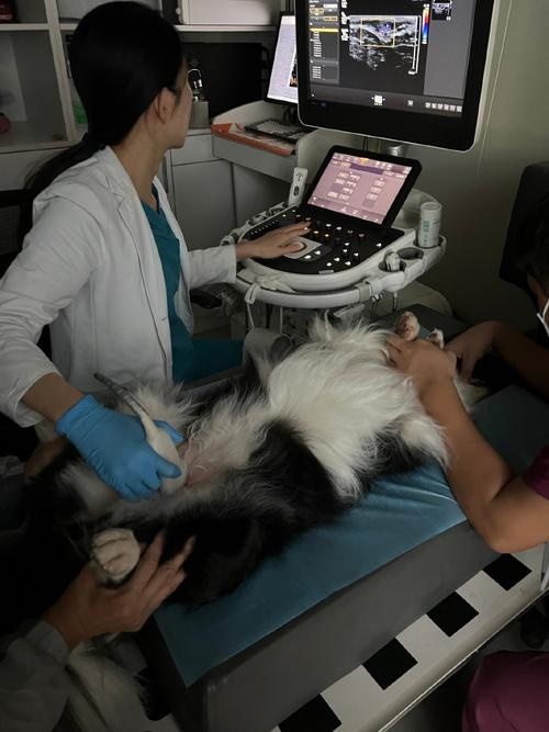
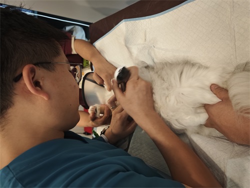
Moreover, portable ultrasound machines are widely used in الخدمات البيطرية المتنقلة, allowing cardiologists to perform complete heart scans even in the pet’s home environment.
Why Ultrasound is Indispensable in Canine Heart Health
Veterinary ultrasound has numerous advantages when it comes to canine cardiology:
-
Non-invasive and low-risk: No sedation is required for most scans.
-
Real-time imaging: Enables immediate clinical decisions.
-
Early detection: Identifies structural and functional issues before clinical symptoms arise.
-
Quantitative assessment: Enables measurement of chamber dimensions, wall thickness, and flow velocity.
By enabling earlier and more precise diagnoses, cardiac ultrasound significantly improves treatment outcomes و life expectancy in dogs with heart disease.
استنتاج
Ultrasound technology has become the gold standard for evaluating canine heart structure and function. It offers detailed, non-invasive insight into left and right ventricular performance, valve motion, chamber size, and blood flow dynamics. As cardiac diseases in dogs continue to be a major health concern globally, ال role of ultrasound in early detection, ongoing monitoring, and informed treatment decisions is more vital than ever.
Veterinarians across the world—especially in Western countries—have embraced cardiac ultrasonography not only for its diagnostic power but also for its practicality and safety. As more pet owners become aware of heart health risks in dogs, routine echocardiographic screening is likely to become a cornerstone of small animal medicine worldwide.
Reference Sources:
-
Thomas, W. P., et al. (1993). Recommendations for standards in transthoracic two-dimensional echocardiography in the dog and cat. Journal of Veterinary Internal Medicine.
-
American College of Veterinary Internal Medicine (ACVIM). (2023). Guidelines for the diagnosis and treatment of canine heart disease.
-
Cornell University College of Veterinary Medicine. (2024). “Echocardiography in Dogs.” https://www.vet.cornell.edu
-
BXL Veterinary Imaging. (2024). “Ultrasound Systems for Veterinary Cardiology.” https://www.bxl-vet.com