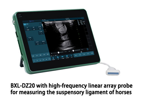
Ultrasound is a vital diagnostic tool in equine medicine, especially for assessing soft tissue injuries like those involving the suspensory ligament. The suspensory apparatus plays a crucial role in supporting the fetlock joint during motion and weight bearing, and any injury to it can be career-ending if not properly diagnosed and managed. This guide will walk you through the professional process of using ultrasound to evaluate the suspensory ligament in horses, from preparation to interpretation.

Understanding the Suspensory Ligament
The suspensory ligament is a strong, fibrous structure that runs down the back of the cannon bone in both the front and hind limbs. It originates from the proximal metacarpus or metatarsus and divides into medial and lateral branches, which insert on the proximal sesamoid bones. In sport horses, particularly jumpers and dressage horses, the suspensory ligament is prone to strain injuries, especially in the hind limbs.
Common signs of a suspensory injury include:
-
Subtle or intermittent lameness
-
Performance issues or reluctance to work
-
Swelling along the cannon bone
-
Pain or reactivity to palpation in the suspensory region
Ultrasound allows for detailed visualization of the ligament’s internal fiber pattern, size, and any lesions that may be present.

Equipment and Preparation
To perform a high-quality ultrasound examination, you’ll need the following:
-
A high-frequency linear ultrasound probe (7.5–12 MHz)
-
Clipping equipment (to remove hair for better probe contact)
-
Ultrasound gel or alcohol
-
Clean towels and gloves
-
A restraining handler or stocks for safety
Clip the hair from the horse’s limb, typically from the level of the carpus/tarsus to the fetlock. Clean the area thoroughly with alcohol to remove dirt and debris, then apply plenty of ultrasound gel to ensure good contact and image clarity.
Positioning the Horse
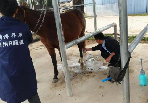
The horse should be standing squarely on a flat surface, bearing weight evenly. It’s critical that the limb is in a normal, weight-bearing position to ensure accurate imaging and measurements. A relaxed, cooperative horse improves the quality of the exam, so allow time for the horse to settle.
For forelimbs, stand facing the limb from the side. For hind limbs, always stand to the side or slightly behind the limb to avoid potential kicking.
Ultrasound Technique
Begin the ultrasound examination by identifying anatomical landmarks. Start proximally at the origin of the suspensory ligament, just below the carpus (or hock in the hind limb), and work distally toward the sesamoids.
Examine the ligament in both transverse (cross-sectional) and longitudinal (lengthwise) planes:
-
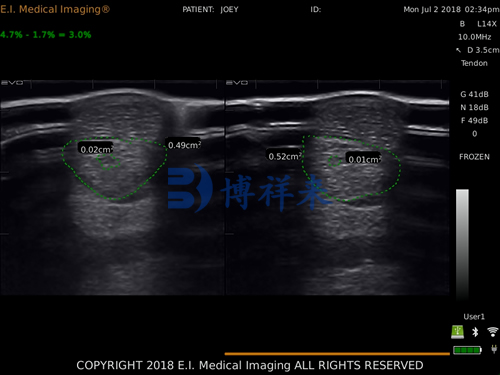
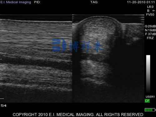
In transverse views, you should see a homogenous, oval to round structure with consistent echogenicity.
-
In longitudinal views, the ligament should show parallel, uniform echogenic lines.
Scan the entire length of the ligament, taking systematic images every 2-3 cm. Pay close attention to any changes in shape, echogenicity (brightness), or the presence of hypoechoic (dark) or anechoic (black) regions that might indicate fiber disruption, hemorrhage, or edema.
Identifying Suspensory Injuries
Injuries to the suspensory ligament are typically classified into three main categories:
-
Proximal Suspensory Desmitis (PSD): Often seen in the hind limb, particularly in sport horses. The ligament may appear enlarged, with areas of reduced echogenicity at the origin.
-
Body Lesions: These are often focal tears or fiber disruption within the mid-body of the ligament. You’ll see an area with loss of the normal fiber pattern and decreased echogenicity.
-
Branch Injuries: The medial and lateral branches may show focal swelling, fluid accumulation, or partial tears.
Quantify the injury by measuring the cross-sectional area (CSA) of the ligament at the lesion site and compare it to the contralateral limb. A greater than 10–15% increase in CSA can be significant, especially in athletic horses.
Common Mistakes to Avoid
-
Inadequate clipping or poor probe contact can degrade image quality.
-
Failure to scan the entire ligament may result in missed lesions.
-
Not comparing both limbs may lead to misinterpretation of mild or early injuries.
-
Misidentifying nearby structures such as the superficial digital flexor tendon or check ligament.
Consistent technique, proper patient handling, and attention to detail are crucial to accurate assessment.
Documenting and Interpreting Results
Always record and store clear images from key locations along the ligament for future reference and monitoring. Include both transverse and longitudinal images of:
-
The origin
-
Mid-body
-
Branches
-
Any abnormal areas
Veterinarians typically score lesions based on echogenicity (grade 0–3) and the percentage of cross-sectional area affected. Follow-up ultrasounds at regular intervals (e.g., every 30–60 days) are used to track healing and adjust the rehabilitation plan.
Treatment and Rehabilitation Planning
The ultrasound findings directly inform the treatment approach, which may include:
-
Rest and controlled exercise
-
Anti-inflammatory medications
-
Shockwave therapy or regenerative therapies (PRP, stem cells)
-
Surgery in chronic PSD cases (e.g., neurectomy and fasciotomy)
Rehabilitation programs often span several months, gradually increasing activity as ultrasound shows improved fiber alignment and reduced lesion size.
استنتاج
Ultrasounding the equine suspensory ligament is an essential diagnostic skill for any equine veterinarian or experienced horse owner involved in performance horse care. With a methodical approach, proper equipment, and a sound understanding of ligament anatomy, ultrasound provides detailed and dynamic insights into injury severity, healing progression, and return-to-work planning.
Early and accurate diagnosis is key to a successful outcome. By mastering the use of ultrasound for suspensory evaluation, you’ll enhance your ability to manage lameness issues effectively and prolong your horse’s athletic career.
