Ultrasound Examination Methods for Boars Using a Porcine Swine Ultrasound System
Porcine Swine ultrasound is one of the primary methods for assessing boar reproductive performance. Visualizing the testicles, epididymis, and accessory gonads is useful for determining the causes of infertility and diagnosing boar reproductive tract diseases. Porcine Swine ultrasound examination of the boar’s reproductive tract must be performed with the boar in an upright position and requires transurethral introduction into a restraining pen.
High-Definition Porcine Swine Ultrasound System
A linear probe is required for transrectal scanning of the accessory gonads. For the testicles, epididymis, and spermatic cord, a convex or convex probe is the preferred device. Frequencies of 5.0–9.0 MHz are used. Lower frequencies are more suitable for tissues requiring greater penetration depth (i.e., the testicles and epididymal body), while higher frequencies are suitable for relatively small structures such as the epididymal head and tail. Therefore, the optimal probe and frequency will depend on the objective of the examination.
Examining the Boar Gonads Using a Linear Array Probe
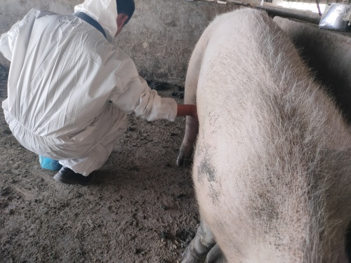
Before scanning the testicles, epididymis, and spermatic cord, the scrotal surface should be cleaned and shaved if necessary. The testicles can be scanned longitudinally or transversely.
Examining boar testicles with ultrasound
Transverse imaging is necessary to determine testicular circumference. Healthy testicular tissue is mesoechoic and has a homogeneous echotexture, with a hyperechoic testis in the center of the testicle (see image below).
Examining boar testicles with ultrasound
Ultrasound images of boar testicles from transverse (A) and longitudinal (B) scans. The testicle is imaged as a hyperechoic spot in the center of the testicle (solid arrow). The boar testicular parenchyma appears mesoechoic and has a homogeneous echotexture. Transverse imaging also provides optimal visualization of the epididymal corpus epididymis (dashed arrow), which typically has a similar ultrasound appearance to the testicle.